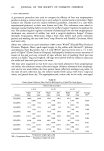
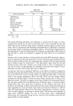
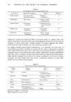
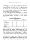
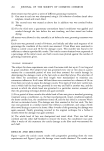
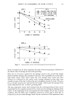
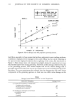
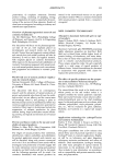
PERSPECTIVES ON AXILLARY ODOR 199 various organisms found in the axilla. The sites were then occlusively covered for 24 hours to ensure multiplication of the organisms. The odor was then assessed organoleptically by three blind observers. Both types of diphtheroids produced intense odor in every instance where their numbers reached 10,000/½m 2. When resident cocci (S. epidermidis and S. saprophyticus) were incubated with apocrine secretion, the acrid apocrine odor was not produced. Instead, a sweaty, acid odor was generated similar to that of isovaleric acid whose presence was demonstrated in a separate study. Gram negatives created odors quite distinctive from either of the above odors. Magnesium- omadine, an antibacterial, reduced bacterial growth and thus all bacterially derived odors. Additionally, some diphtheroids after repeated subcultivation failed to generate strong odors on incubation with apocrine secretion. This explains lack of odor in some cases with good bacterial growth. Jackman has also confirmed the presence of two distinct types of axillary flora with the coryneforms being higher in males and contributing the more pronounced body odor (22b). BIOCHEMICAL ASPECTS OF AXILLARY ODOR The axillary odor has been described in astonishingly different ways. The characteriza- tions include: goat-like, over-ripe peaches, chlorinared urine, burnt coffee-beans, ammoniared Valerian, etc. These quixotic designations reveal the inadequacy of subjective perceptions and the fact rhar at least two odor types are being described. With the advent of combined gas chromatography-mass specrromerry and radioimmu- noassay techniques ir becomes possible ro investigate the composition of apocrine secretion and ro identify the odor chemicals. Samples of the axillary surface lipids contain considerably more cholesterol and cholesterol esters than surface lipids from other sebum rich areas such as the face (Table V). This reflects the high concentration of free cholesterol in apocrine secretion itself. Various sreroidal substances have been identified in samples from the axilla, usually collected in pads held in the axillary vault (Table VI). The two A•6-androgen steroids, 5o•-androsr-16-en-3-one, a kerone, and 5o•-androsr-16-en-3o•-ol, an alcohol, together account for parr of the acrid axillary odor. The latter has a musky odor and is nor altogether unpleasant (Table III). The kerone confers the disagreeable and Table V Axillary and Apocrine Lipid Profiles Percent Composition Glandular Secretion Apocrine • Sebaceous 2 Extract Skin Surface Axillae Facial Cholesterol 76.2% 3.4% 8.9% 1.5% Cholesterol Esters 0.9%* 21.8% 8.8% 3.0% Wax Esters 3.6%* 21.2% 26.0% Squalene 0.2%* 19.0% 13.4% 12.0% Glycerides and Fatty Acids 19.2%* 55.9% 47.4% 57.5% Total Lipid 20 3tg/3tl -- 60 3tg/cm 2 100 3tg/cm 2 Protein 90 3tg/3tl -- -- -- •Stimulated and collected at skin surface •Collected from microdisection of gland (39) *Probably of sebaceous origin

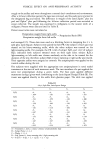
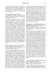
200 JOURNAL OF THE SOCIETY OF COSMETIC CHEMISTS Table VI Steroids Found in Human Axillae Steroid Sample Reference Axillary Hairs and sweat 40 Androst-4-ene-3, 17-dione Androsterone (sulphate) DHA (sulphate) Cholesterol Androst-4-ene-3, 17 dione Pregn-5-en-3/•-ol-20-one 5c•-androst-16-en-3c•-ol 5c•-androst-16-en-3-one Androsterone (sulphate) DHA (sulphate) Cholesterol Axillary Sweat 41 Axillary Sweat 42 Axillary Sweat 24, 43 Apocrine Secretion 25 dominant odor and in odor description studies has been labeled urine, sweaty, perspiration, or animal (23). Recently investigators utilizing radioimmunoassay tech- niques have demonstrated differences in concentration of androstenone in male (3-310 ng) and female (3.5-11 ng) subjects (24). These results correlate well with the overall sex differences in bacteriology discussed above. In pure apocrine secretion we have demonstrated the presence of two sulfated steroids, androsterone sulfate and dehydroepiandrosterone sulfate (25). Whether either of these are precursors of the odorous steroids is unknown (26). The cholesterol, steroids, and proteinaceous (10%) substances present in apocrine secretion provide a unique substrate for bacterial growth and odor development. Other substances in the axilla originating from the sebaceous and eccrine glands may contribute indirectly to the total odor profile. Sebum intermingles with apocrine secretion in the follicular infundibulum and contains about 10% squalene, a material which fragrance formulators use as a "fixative" to make the odor more durable. Interestingly, the sebaceous gland of the musk deer produces many androgen steroids in addition to lipids and the odorous muscone (27). That secretion is valued both in perfumery for its musk odor and as an important drug for its various pharmacological effects. Finally, in vitro incubation of apocrine secretion and micrococci produces a sweaty odor which has been identified as isovaleric acid by odor concentration and analysis by gas chromatography-mass spectrometry (44). A similar incubation of apocrine secretion with diphtheroids produces the typical acrid odor, though fresh bacterial isolates are needed to obtain consistent results. Isovaleric acid was detected in the gas chromato- graphic profile but the main odor components, presumably steroids, have not as yet been characterized in these cultures. In the case of these steroids, particularly androst-16-en-3-one, the sensitivity of the much underrated human nose exceeds that of the instrument. SUMMARY: The study of axillary odors presents an interesting paradox. Knowledge of the responsible bacteria and an understanding of their interaction with apocrine secretion will lead to alternative and possibly better methods of odor control. It is clear we are dealing with two bacterial populations and consequent differences in odor profiles.
Purchased for the exclusive use of nofirst nolast (unknown)
From: SCC Media Library & Resource Center (library.scconline.org)