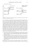
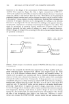
232 JOURNAL OF THE SOCIETY OF COSMETIC CHEMISTS streptomycin sulfate (Sigma) were placed in 150 X 25-mm petri dishes and dried overnight at 35øC prior to inoculation. The convex surface of the agar plates was streaked with the diluted cultures and allowed to air dry for 30 minutes before attaching to panelists' forearms. CULTURES The bacterial cultures were grown overnight in AATCC bacteriostasis broth (Difco) at 35øC. The third to seventh consecutive overnight cultures were used to prepare the diluted inoculums for each experiment. (A) Staphylococcus epidermidis ATCC 155 was maintained on trypticase soy agar (BBL). A 4-mm loopful of an appropriate dilution of a 23-hour-old culture was streaked on the dried agar plates to obtain the desired number of colony-forming units (CFU) per plate. (B) Staphylococcus epidermidis JEH 51 was maintained on trypticase soy agar containing 1 mg/ml streptomycin sulfate. This culture was isolated as a spontaneous streptomycin- resistant colony from S, epidermidis ATCC 14990 and has the same agar media growth rate and biochemical properties on Staph-Indent Strips (API) as the parent strain. A 4-ram loopful of the diluted 23-hour-old culture in 0.1% peptone broth was streaked on trypticase soy agar containing 1 mg/ml streptomycin sulfate to obtain approximately 1000-2000 CFU per plate. PROCEDURE Six to eight panelists were used for comparing two soap bars in each test. The panelists were instructed to use a placebo soap for all bathing and washing of hands and forearms for 1 week prior to the test. On test days supervised washings of the forearms with the randomly assigned soap bars were conducted in the laboratory. Three agar plates streaked with S. epidermidis ATCC 155 were attached to the volar surface of the washed forearms with Blenderm © Surgical (3M) or Dermilite II © (Johnson & Johnson) tape for either 2 or 4 hours. For later experiments trypticase soy agar + 1 mg/ml streptomycin sulfate plates streaked with JEH-51 were taped to the volar surface of the forearms with 1" by 3" strips of Microfoam © tape (3M) and wrapped with an Ace © type elastic bandage (Becton-Dickinson) for 30 minutes. • The plates were removed, placed in 150 x 25-mm petri dishes, and incubated at 35øC. Control plates were not attached to panelists' forearms but were placed directly in the 35øC incubator. After 24 hours, colonies were counted using a 10 x dissecting microscope. The regimen for the supervised laboratory washings consisted of washing the hands with the placebo soap bar, wetting the volar surface of the forearms and the assigned soap bar under the tap, rubbing the soap bar to the volar surface of the forearm for 15 seconds, lathering for 30 seconds, rinsing under tap water for 30 seconds, blotting forearms dry with paper towels, and repeating the whole procedure for the other forearm and soap bar. In all the experiments performed there was at least a 2-hour drying out time between the different washes conducted over a 2 to 4 day period. • All three taping methods held the entire inoculated portion of the agar plates firmly to the panelists' forearms. However, the Microfoam strips were the easiest and least painful to remove from panelists' skin.

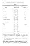
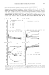
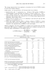
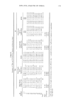
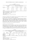
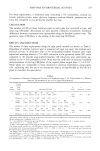
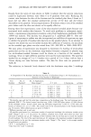
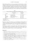
SOAP BAR ANTIBACTERIAL ACTIVITY 233 For these experiments, a deodorant soap containing 1.5% triclocarban, sodium tal- lowate, sodium cocoate, water, glycerin, fragrance, sodium chloride, preservatives, and color was compared to non-germicide placebo bar soap. CALCULATION The number of CFUs (colony forming units) on each plate was converted to logs, and mean log CFU/plate determined for each panelist's forearms (treatment). Statistical differences between treatments were determined using the Student's paired t-test. The geometric mean CFU/plate is the antilog of the mean log CFU/plate. RESULTS AND DISCUSSION The results of three experiments using the agar patch method are shown in Table I. Regardless of whether nutrient agar or mannitol salt agar was used, the residual anti- bacterial activity of deodorant soap (1.5% triclocarban)-washed forearms after eight supervised washings showed a 79 to 88% reduction in the geometric mean CFU count compared to the placebo soap-washed forearms. All reductions were statistically sig- nificant at the •.025 probability level. Note that the coefficient of variation (standard deviation/mean) of the log CFU/plate of the control plates ranged from 3.3 to 6.4%. These values are comparable to those obtained in previous experiments using pipets (10), indicating that the use of an inoculation loop is as reproducible as the use of a pipet to inoculate the agar plates. Table I A Comparison of Residual Antibacterial Activity of a Deodorant Soap (1.5 % Triclocarban) vs. a Placebo Soap (no Triclocarban) by the In Vivo Agar Patch Method • Panehst Initials/ Test Soap • % Number Used Mean Log CFU/Plate + S.D. Reducnon c p-Value d Imt•als LS RD WF PF RB RC Placebo 1.9318 1.8949 2.3514 2.3784 1.9898 1.8647 .6874 79.5 .005 ñ .3165 Deodorant t.1t27 1.7703 1.3809 1.7807 1.0249 1.2173 Initials RL RD PF PW BB GG CS MH Placebo 1.0887 2.2535 2.2503 2.5692 2.087 1.8221 1.5519 1.8345 .732 81.5 .025 ñ .7120 Deodorant 1.1937 1.5576 0.8844 2.2329 0 1.6806 1.t032 0.9488 Initials GG CS SE JM PF RB MH RC Placebo 1.2915 1.2896 1.1286 1.7654 1.9211 1.7472 1.4053 1.5853 .9401 88.4 .001 q-.3916 Deodorant 0.477t 0.7101 0.6990 0.6667 0.2817 1.0195 0.4407 0.318t • All panehsts washed with the test bars for 8 applications. The placebo soap had a d•fferent base composition from the deodorant soap. Th_e test bacterium was S. epidermzdit ATCC t55. b d = mean difference of the placebo _mean log CFU/plate--the deodorant mean log CFU/plate. S.D. = standard deviation. • The % reduction = (1 - 1/antilog d) 100. d p-value = the smallest alpha level at which the null hypothesis of no differences can be relected. e The desired control inoculum was t00 CFU/plate on nutrient agar using a 1/10,000 dilution of the overnight culture in AATTC bacteriostasis broth. Actual control mean log CFU/plate q- S.D. = 2.0188 q- 0.1137 (n = 7). The contact time was 4 hours between the washed forearms and the inoculated plate. f The desired control inoculum was 200 CFU/plate on mannitol salt agar using a 3/10,000 dilution in 0.1% peptone. Actual control mean log CFU/plate q- S.D. = 2.2703 ñ . 1460 (n = 12). The contact t•me was 4 hours. Because some of the test plates contained no surviving CFU, "1" was added to all CFU/plate counts to allow mean log CFU/plate to be calculated. g The desired control inoculum was 400 CFU/plate on manmtol salt agar using a 6/10,000 dilution in 0.1% peptone. Actual control mean log CFU/plate q- S.D. = 2.4642 q- .08t0 (n = 8). Contact time = 2 hr.
Purchased for the exclusive use of nofirst nolast (unknown)
From: SCC Media Library & Resource Center (library.scconline.org)