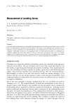
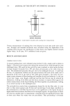
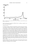
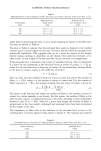
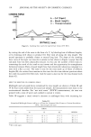
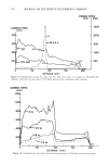
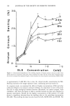
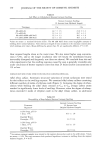
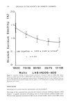
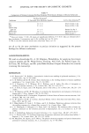
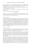
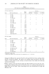
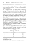
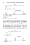
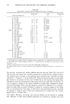
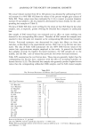
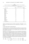
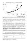
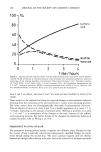
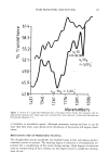
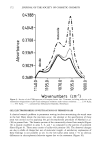
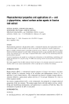
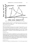
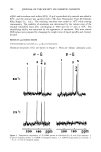
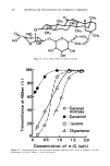
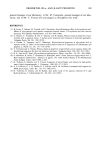
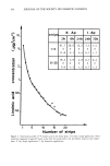
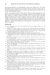
164 JOURNAL OF THE SOCIETY OF COSMETIC CHEMISTS 100- 50- I I I ! •" 5 10 15 20 [ mini BI ec[ching lime Figure 2. Oxidative treatment on isolated melanin. Correlation between oxidized disulfide bridges in % ( ..... ) and loss of weight of the granules in % ( ) (Giesen (3)). A method for direct measurement of cystine oxides in hair is via infrared spectroscopy. Fourier transform infrared spectroscopy (FTIR) offers the possibility of demonstrating the existence of cystine oxides and cysteic acid non-destructively in one step by ob- serving the sulfur-oxygen vibrations. Application of this technique to the investigation of oxidative damage on human hair has already been described in the literature (16). Schumacher-Hamedat found during her work on this subject an increase in monoxide and dioxide concentration after per- formic acid oxidation of human hair. Using peroxide-containing bleaching solutions, however, only the monoxides are found (Figure 5) these findings are identical to those obtained in studies of the model compound ACM (17). The oxides even appear in untreated hair, as does cysteic acid, presumably due to weathering. TWO-DIMENSIONAL GEL ELECTROPHORESIS OF UNTREATED AND BLEACHED HAIR SAMPLES The application of two-dimensional gel electrophoresis techniques in the field of human Table III Cysteic Acid Values (mol%) in Total Hydrolysates of Hair in Two Different Media (FiShles et al. (13)) 6N-HC1 + 3 Vol% Hair Sample 6N-HC1 Thioglycollic Acid Untreated 0.60 0.44 47 h irradiation • 0.73 0.51 126 h irradiation • 0.81 0.58 Oxidized • 3.22 2.70 Xenotest apparatus Model 150--Filter system 6 IR/1 UV. Bleached under unknown conditions in a hair dresser's salon.

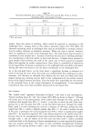
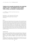
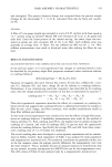
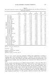
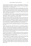
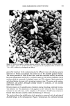
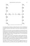
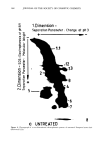
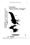
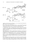
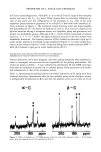
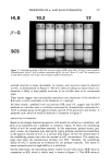
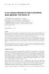
HAIR BLEACHING AND WAVING 165 oeH 3 NH NH oe0 HE NH oe0 I oeH• [H 2 S S [H 2 EO I EH I NH I EO EH 3 Figure 3. N,N'-bisacetyl-L-cystine-bismethylamide (ACM). hair research allows the separation of unfractionated protein extracts. Radiolabeling of the investigated keratins increases the detection sensitivity so that very small samples are sufficient, allowing for high resolution of the protein components during electro- phoresis. The first step of the procedure is the reductive solubilization of the keratins followed by the alkylation of the cysteine residues thus obtained with iodo(2-•4C)-acetic acid. The separation parameter for the first dimension is the charge of the proteins, whereas in the second dimension it is the apparent molecular weight in the presence of sodium dodecyl sulfate (SDS). Proteins are located by fluorography (Figure 6). For full experimental details, refer to Marshall et al. (18). After investigation of more than one hundred different single hair samples under iden- tical conditions, it was found that a basic pattern was always repeated (18-20). How- ever, different exogenous or endogenous influences cause variations. Fluorographs showing the changes in patterns obtained with untreated (Figure 6) and bleached human hair (Figure 7) illustrate some of the possibilities of this technique for detecting the chemical changes that occur during the cosmetic process. The changes in the fluorographs of the bleached samples compared with those of un- treated hair are: Area 1.1 is weaker, areas 1 and 2 are unchanged with regard to the molecular weight of the proteins but are obviously modified with respect to the charge,
Purchased for the exclusive use of nofirst nolast (unknown)
From: SCC Media Library & Resource Center (library.scconline.org)