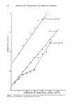
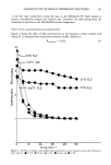
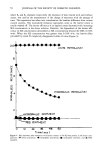
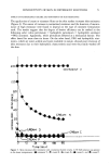
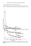
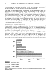
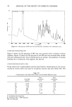
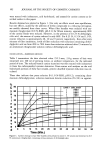
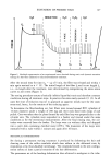
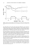
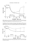
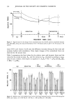
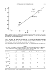
j. Soc. Cosmet. Chem., 38, 63-75 (March/April 1987) The effect of barrier creams on the electrical conductivity of excised skin during exposure to detergents j. c. LIU, M. J. HUANG, Y. SUN, and Y. W. CHIEN, College of Pharmacy, P.O. Box 789, Rutgers--The State University of New Jersey, Piscataway, NJ 08854. Received February 26, 1986. Synopsis Electrical conductivity (K) was measured in excised hairless mouse skin after treatment with selected skin creams or cream ingredients. The measurements were performed with the skin samples mounted in modi- fied diffusion cells which contained sodium lauryl sulfate (SLS) or other detergent solutions in the donor compartment. The receptor compartment was filled with physiological saline. Analyses of the conductivity values as a function of time post-exposure to detergent show that this experimental design might be useful for studying the effects of barrier creams on epidermal permitivity and the removal of the effect by deter- gents. White petrolatum and a commercial cream containing white petrolatum were found to be the most effective in decreasing electrical permitivity and in resisting removal by SLS. Barrier creams based on hydrophilic substances and little or no hydrocarbon ingredient were found to be less effective. INTRODUCTION Repeated exposure of human skin to SLS and other detergents causes a progressive loss in the electrical resistance of stratum corneum (SC), and clinical signs of skin damage such as chapping and erythema (1,2) appear. According to a study by Serban et al. (2) the loss in electrical resistance precedes erythema, thereby indicating that an impair- ment in the epidermal barrier function must occur before dermal tissue becomes in- volved. Treatment with barrier creams prior to exposure to SLS delays the loss of elec- trical resistance of epidermis and produces lower erythema scores (2). The most plau- sible explanation for the effect is that lipophilic ingredients from the lotions partition into the stratum corneum. There they act as a temporary barrier which impedes the penetration of the detergents and, thus, confer some protection to the skin. In order to systematically characterize the barrier function of the skin and its response to topical treatment, an in vitro electroconductivity method was developed in contrast to re- ported in vivo methods (3-8). In the present experiments, we applied various skin creams to freshly excised hairless mouse skin. After a period of equilibration, the treated sites were kept under detergent solutions and the electrical conductivities of the samples were monitored over a period of 60 minutes and longer. We report that with some treatments there occurs an initial increase in resistance of 100 to 200-fold. With the passage of time, the resistance falls gradually to near control values. Preliminary data reported here suggest that the experimental design may be useful for studying the effects of barrier creams on the resistance of epidermis to detergent damage. 63

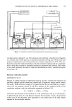
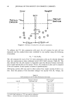
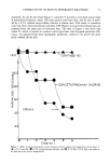
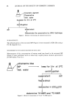
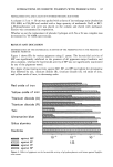
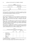
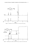
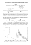
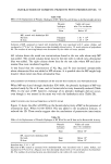
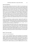
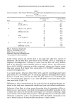
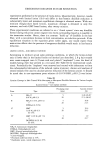
64 JOURNAL OF THE SOCIETY OF COSMETIC CHEMISTS EXPERIMENTAL MATERIAL AND EQUIPMENT Sodium lauryl sulfate, cetylpyridinium chloride (Fisher Chemicals, Springfield, NJ), Tween 80 (polysorbate 80 USP, Ruger Chemical Co., Inc., Irvington, NJ), commercial creams (Table I), and white petrolatum NF were used as purchased. Ointment bases: PEG ointment USP (40% polyethylene glycol 3350 and 60% polyethylene glycol 400), hydrophilic ointment USP, and hydrophilic petrolatum USP were prepared in the labo- ratory following the formulations and methods listed in the compendia (9). Hydrodynamically calibrated Valia-Chien diffusion cells (10) (Crown Glass Company, Somerville, NJ) and a conductometer with a frequency range of 73 to 50,000 Hz (Model CDM 83, Rainin Instrument Co., Inc., Ridgefield, NJ) were connected in parallel through a cell selector switch (Figure 1). Six wire-type platinum (0.5 mm in diameter, Alfa Products, Danvers, MA) electrodes were prepared. They were installed in the diffusion cells, replacing the glas s stopcocks on both sides of the skin. The con- ductometer completed the circuit with one of the diffusion cells, as the cell selector was switched to it. SKIN PREPARATION Full-thickness skin freshly excised from 5-7-week-old male hairless mice (HRS/J strain, Jackson Laboratories, Bar Harbor, ME) was used. The hairless mice were each sacrificed by cervical dislocation. The whole-thickness abdominal skin (ca. 3 x 3 cm 2) was excised from the animal and the excess subcutaneous tissue was removed (11). EFFECT OF BARRIER CREAMS ON THE ELECTRICAL PERMITIVITY The skin samples were each mounted on the receptor compartment of the V-C skin permeation system with the stratum corneum surface facing up. A test cream or oint- ment formulation was then applied onto the stratum corneum with a spacer to give a constant thickness of 0.15 - 0.01 mm and allowed to air dry at ambient conditions for one hour. The donor compartment was placed on the top of the cream-skin laminate and the whole skin permeation system was then assembled horizontally. An aqueous solution with a known concentration of a detergent was added into the donor compart- ment and saline solution was filled into the receptor compartment. The platinum electrodes were immersed into the donor and receptor solutions ca. 5.5 Table I Commercial Barrier Creams Evaluated Product Ingredients Cream C Cream D Cream N Cream P Cream V Beeswax/mineral oil/polyoxyethylene sorbital lanolin/water Mineral oil/water/cetearyl alc./glycerin/sodium lauryl sulfate Water/mineral oil/petrolatum/wax/lanolin alcohol/paraffin/magnesium sulfate/decyl oleate, octyl dodecanol/etc. Mineral oil/cetyl alc./water/stearic acid/potassium hydroxide White petrolatum
Purchased for the exclusive use of nofirst nolast (unknown)
From: SCC Media Library & Resource Center (library.scconline.org)