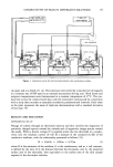
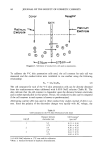
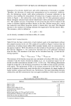
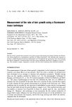
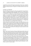
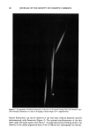
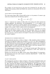
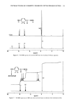
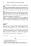
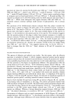
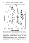
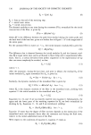
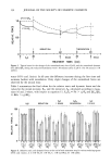
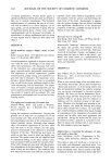
FTIR OF SWEAT GLANDS & ALUMINUM SALTS ! 11 eter purged with dry nitrogen and equipped with a hot wire source and a triglycine sulfate detector. Spectra were recorded at 4 cm-• resolution with the use of boxcar apodization, and 1000 scans were signal averaged for each spectrum to attain a signal- to-noise ratio yielding good spectral subtraction conditions. Two microsampling techniques were used for all studies with modifications when needed. A 6X beam condenser was used to concentrate the beam to about 2 mm diam- eter at the focal point. The first technique utilized a high pressure diamond anvil cell (High Pressure Diamond Optics, Tucson, AZ) in which the sample was pressed be- tween the two polished diamond faces. The diamonds, mounted on pistons, were squeezed by the lever action of a spring on the pistons. The pressure was cycled to spread the sample, and spectra were recorded at zero pressure with the spring loosened. A full description of the diamond cell is given elsewhere (9). Where larger samples were available, they were flattened between two gold mesh screen grids (of the type used for electron microscopy) and masked on the periphery with larger (1 x 2 mm) crossed grids. The grids and sample were mounted in a specially fabricated microsample holder, which likewise held the sample at the focal point of the beam condenser. Reference spectra were generated with either a clean diamond cell or empty grids in the 6X beam condenser. Sample holes in the infrared windows were masked to minimize spectral dilution or fringing effects. ISOLATION OF HUMAN ECCRINE SWEAT GLANDS Biopsies (!) 2 mm) were taken from the volar forearms of two human males. One forearm was treated overnight with an occlusive patch of 20% ACH the other arm had no treatment. The following day the subjects were thermally stressed at 100øF and 35% RH and biopsies taken at sites devoid of sweating. The biopsies were preserved under liquid nitrogen and brought to room temperature for dissection. They were fixed for two days at 4øC in 6.25% glutaraldehyde buffered with 0.1M Sorensen's phosphate buffer, pH 7.3. Each biopsy specimen was sliced with a razor blade into 0.2-mm sections parallel to several sweat glands. Immersion in glycerine revealed the gross anatomy of the sweat gland with a ductal area substantially darker (under transmitted light) than any surrounding tissue. The dark material was considered to be the region of poral occlusion or a plug from an ACH-inhibited sweat duct. Sections were also cut to demonstrate sweat ducts from the untreated forearm where no dark material was found. A sliced section is shown in Figure !. The dark plug material (usually found near the stratum corneum/viable epidermis interface) was gently removed from the surrounding tissue with specially ground, fine-tipped tweezers. It was not possible to remove cleanly the dark plug material without drawing a small amount of surrounding tissue with it. Approximately three or four excised plugs were used to compose each of the samples designated as plug # ! and #2 and taken from different subjects. Additional stratum corneum was dissected from the same ACH-treated biopsy as plug #1 but about 2 mm away from the sweat duct. A control sample was sectioned from the biopsy from the untreated forearm and dissected to contain part of the sweat duct as well as surrounding stratum corneum. All FTIR analyses in these experiments were carried out using the high pressure diamond anvil cell. Although the plug samples were thinned considerably in the cell, they were not large enough to completely fill the diamond windows (--1 mm2). The beam was partially masked with an iris to minimize spectral dilution effects. The spectral quality from plug #2 was superior to that from

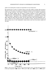
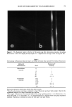
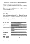
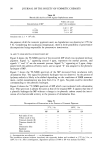
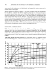
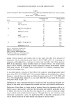
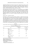
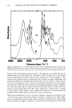
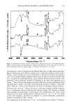
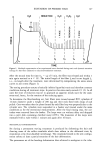
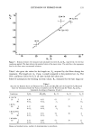
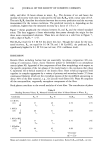
112 JOURNAL OF THE SOCIETY OF COSMETIC CHEMISTS Figure I. Light photomicrograph of thin section through ACH-inhibited sweat glands. Dark areas of poral occlusion, arrows, appear near stratum corneum/viable epidermal interface. -- 100 X. plug # 1. About 70% of the area of the diamonds was covered in plug #2 and about 25% in plug # 1, so spectral dilution effects were smaller in plug #2. Since the spectra were different, they will be considered separately. RELATED PROTEINS AND PRECIPITATES To aid in characterizing the protein portion of the plug spectra, we examined the four sweat proteins described in Table I in the diamond cell. A portion of the human sweat concentrate was redissolved in water and combined with a 20% ACH solution, forming a precipitate. This precipitate was filtered, air dried, and also analyzed in the diamond cell. These experiments were performed with complete coverage of the diamond windows by the sample. A 5% human albumin solution was combined with a 20% ACH solution. The spec- trum of the precipitate thus formed proved to be albumin (an exact match of Figure 3B, but not shown) because of a salting out process. When the 5% albumin was combined with a 5% ACH solution, significant spectral changes, indicating interaction, appeared in the dried precipitate. All of the resulting solutions were close to the physiologic pH of 6.0. Similar attempts to form precipitates from a 5% glycoprotein solution, with either 20% or 5% ACH solutions, were unsuccessful. Dried films from these solutions were analyzed but the resulting spectra were dominated by water and significantly different from any others in the study. These results indicated that ACH-glycoprotein complexes were not plug formers. IN VITRO ACH-TREATED STRATUM CORNEUM A variety of in vitro experiments was performed on biopsies of human stratum corneum
Purchased for the exclusive use of nofirst nolast (unknown)
From: SCC Media Library & Resource Center (library.scconline.org)