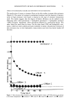
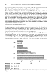
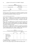
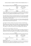
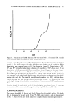
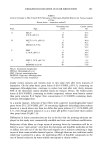
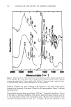
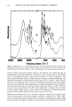
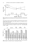
84 JOURNAL OF THE SOCIETY OF COSMETIC CHEMISTS MATERIALS AND METHODS COSMETIC PIGMENTS The following pigments were used: red oxide of iron, yellow oxide of iron, titanium dioxide (Rutile), titanium dioxide (Anatase), talc, ultramarine blue, kaolinite, and silica alumina. These cosmetic pigments were Japanese cosmetic-grade materials. PRESERVATIVES Methyl p-hydroxybenzoate (MP), ethyl p-hydroxybenzoate (EP), propyl p-hydroxyben- zoate (PP), and butyl p-hydroxybenzoate (BP) were used. They were Japanese Pharma- copoeia reagent grade. TEST ORGANISMS Staphylococcus aureus 209P, Escherichia coli o-1, Pseudomonas aeruginosa IAM 1007, and a laboratory strain of Candida albicans were used in this study. CULTURE MEDIA Bacteria were grown on nutrient agar plates and yeast was grown on potate dextrose agar plates. Cells in the late phase were used for inoculations. DETERMINATION OF THE BACTERICIDAL ACTIVITIES OF THE PRESERVATIVES IN THE PRESENCE OF COSMETIC PIGMENTS Actively growing bacteria were cultivated on nutrient agar plates for 18 hrs, at 37øC. The growth was aseptically washed off with 5 ml sterile distilled water. The suspensions were freed by agitation from microorganism clumps and used as inocula. Each of the reaction systems consisted of 2.5 ml of the individual solutions of the preservatives, 2.0 ml of the sterile suspension of the material under test, and 0.5 ml of the inoculum (Table I). The pH of these reaction mixtures were not adjusted. The mixture was vigorously shaken and incubated at 37øC or 25øC for 5 hr. Aliquots were then decinormally diluted with a 1% sterile solution of polysorbate 80 (7). Table I Final Concentrations of the Components of the Incubation Mixture Component Final concentration Cosmetic pigment 0.05 g Preservative MP 10 mg EP 8.5 PP 2.5 BP 0.1 Water (Dist.) to 5.0 ml Inoculum size: about 108 cells per test tube.

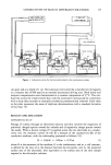
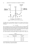
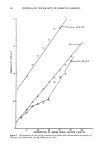
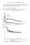
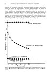
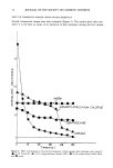
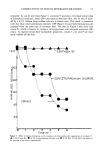
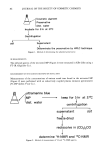
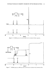
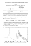
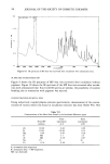
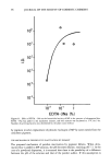
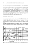
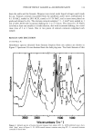
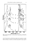
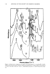
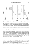
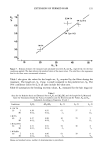
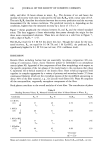
INTERACTIONS OF COSMETIC PIGMENTS WITH PRESERVATIVES 85 The numbers of surviving bacteria and yeast were determined by the plate count method. The necessary controls and blanks were carried out concurrently with the test experiments. CALCULATION OF INACTIVATION DEGREE The inactivation degree (ID) of bactericidal activity in the presence of cosmetic pig- ments was calculated with the following equation: d/c d' a ID - - A x l0 B - b/a b ß c where "a" represents the number of viable cells in the sample tube which contains the cosmetic pigments, the preservative, and microorganism "b" is the number in the blank tube which contains the cosmetic pigment and microorganism "c" is the number in the control 1 which contains the preservative and the microorganism and "d" is the number in the control 2 which contains only the microorganism. When the resulting B is about 0, the bactericidal activity is considered to be little affected by the cosmetic pigment tested. On the other hand, when B is an integral number (ID 1), this indicates inactivation by pigment. Thus the exponential number B in the equation is directly related to the degree of inactivation of the preservative by the cosmetic pigment. DETERMINATION OF ADSORBED PRESERVATIVE The experimental procedure is shown in Figure 1. The incubation mixture (without inoculum) was vigorously shaken and incubated at 37øC for 5 hr. After centrifugation, the solubilized amounts of preservatives in the supernatant solutions were determined by high performance liquid chromatography (HPLC) and compared with the amounts originally added. A model ALC/GPC 201 HPLC (Waters Associates) equipped with a Waters Model 730 data module was used in this study. The chromatographic column (4 mm X 150 mm) was stainless steel. Warm water ran through the jacket to maintain the column temperature at 40øC. The column was packed with Lichrosorb PR-18 (5Ix, Merk). Samples were injected into the HPLC column using a Waters Model 710 autosampler. All experiments were done under isocratic conditions (H20:CH3OH = 20:80). Flow rate was 1.5 ml/min. •H AND 13C-NMR ANALYSIS The experimental procedure is shown in Figure 2. One gram of ultramarine blue and 0.2 g of MP were dispersed in 100 ml of distilled water and incubated at 37øC for 5 hr. After removing the pigment fraction by centrifugation, this supernatant fraction was freeze-dried to recover the solubilized MP. The •H and •3C-NMR spectra of the recovered MP were measured in perdeuteroacetone with tetramethylsilane as internal standard, using a JEOL JMN-FX 100 NMR spectrometer.
Purchased for the exclusive use of nofirst nolast (unknown)
From: SCC Media Library & Resource Center (library.scconline.org)