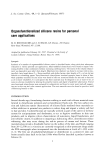
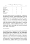
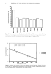
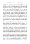
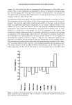
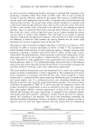
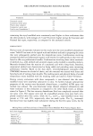
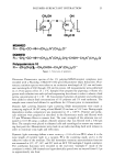
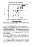
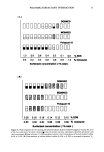
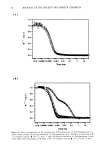
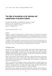
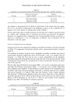
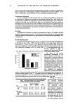
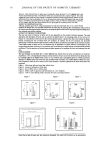
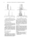
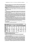
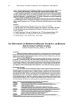
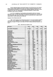
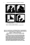
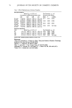
54 JOURNAL OF THE SOCIETY OF COSMETIC CHEMISTS (vIc) Lotion for Sensitive Dry Skin (Chesebrough-Ponds, USA), pH 7.8. Lotions were applied once daily at 3 mg/cm 2 for 3 weeks (Sundays excluded) to designated sites on the back of HGPs. At the end of 3 weeks, HGPs were sacrificed for the in vitro percutaneous absorption studies. Percutaneous Absorption In vitro skin absorption of HQ and musk xylol was measured through HGP skin dermatomed to a thickness of-200-gm and assembled in flow-through diffusion cells. Skin viability was maintained in the diffusion cells during the 24-hr studies by using physiological media. The barrier integrity of untreated, GA- and VIC-treated HGP skin was assessed by determining [3H]water absorption prior to each study (20 rain test•). []4C]HQ or []4C]musk xylol was applied to skin in an oil-in-water emulsion vehicle at a 2% or 0.20% concentration, respectively. At 24 hr, unabsorbed compound was washed from skin with soap and water and the radioactivity recovered in the skin and receptor fluid was determined. Histology Full-thickness skin samples were taken from treatment sites, fixed in 10% formalin, and then stained for light microscopic evaluation. The number of epidermal cell layers was determined from fixed skin sections the thickness of the viable epidermis was measured from photographs (50X magnification). Results and Discussion The effect of GA formulations on skin mover was determined by measuring the disappearance of DC from the skin surface. HGPs were able to tolerate a once-a-day dosing with the GA formulations for 2 weeks, followed by DC application, and then continued daily dosing until fluorescence disappeared. The 5% and 10% GA treatment reduced SC turnover times by 36% and 39%, respectively, compared to the VIC control (Fig. 1). A 3-week pretreatment time was, Fig 2. Glycollc Acid Effect on Stretum Corneum Turnover • 25 e, 20 I-- i• 15 • lO I-- 5 t/ntreltld vllemlne IC s• olycollc Acid 10'/. Olycollc Acid Pretreatment FormulatlonB therefore, sufficient to effect an increase in SC turnover in HGPs used for the skin absorption studies. The effect of the various pretreatment formulations on the barrier integrity of HGP skin was determined by measuring [3H]water absorption through skin prior to the HQ and musk xylol in vitro studies. No significant difference in the percent of the applied [•H]water absorbed was detected between untreated skin and the pretreatment formulations. The percutaneous absorption of HQ was also unaffected by pretreatment applications. No significant difference was found between the partial (receptor fluid or skin) or total absorption values measured for the different treatment groups (Table 1). In general, musk xylol absorption was unaffected by pretreatment applications. However, total absorption values for VIC, 5% GA, and 10% GA were significantly different from untreated skin but were not different from each other. Table 1. In Vitro Skin Absorption of Two Cosmetic Ingredients Following Pretreatment of Hairless Guinea Pig Skin Pretreatment Formulations Untreated VI_•C 5% GA 10%(•A -Q receptor fluid 4.25 + 0.56 5.89 + 0.30 6.44 + 0.88 4.40 4- 0.52 skin 15.0 + 0.67 13.2 + 1.07 15.4 + 1.02 16.1 + 1.82 total 19.3 + 0.43 19.0 + 1.03 21.8 4- 1.87 20.5 + 2.10 Musk Xylol receptor fluid 30.3 + 2.49 23.6 + 0.17 20.7+ 4.23 21.6 + 3.56 skin 18.0 + 2.55 18.8 + 1.21 16.1 + 3.20 18.8 4- 2.12 total 48.3 + 1.73 42.4 4- 1.07 36.7 + 1.31 40.4 + 1.72 Although no increase in penetration of HQ or musk xylol was observed following pretreatment with GA lotions to HGP skin, the structure of skin was drastically altered. Histological examination of the skin revealed a similarity in thickness of untreated and VIC- treated skin (5.0 cell layers) and a thickening of the epidermis in skin treated with 5% GA (9.8 cell layers) and 10% GA (8.1 cell layers). Measurements of the viable epidermal thickness

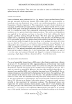
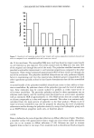
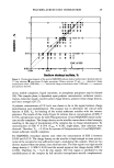
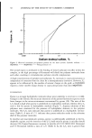
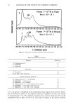
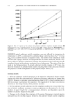
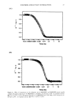
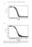
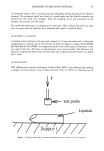
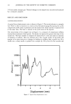
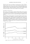
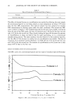
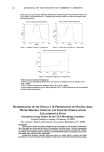
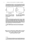
PREPRINTS OF THE 1997 ANNUAL SCIENTIFIC SEMINAR 55 indicated as much as a four-fold increase in thickness compared with untreated and VIC-treated skin. In addition, there appeared to be hypertrophy of the epithelium lining the hair follicles. References 1. Leyden, J. J., Lavker, R. M., Grove, G., and Kaidbey, K. J. Geriatr. Dermatol., 3 (Suppl A), 33A (1995) 2. Lavk'er, R. M.• Kaidbey, K., and Leyden, J. J. J. Amer. Acad. Dermatol., 26, 535 (1992) 3. Jansen, L. H., Hojyo~Tomoko, M. T., and Kligman, A.M. Brit. J. Dermatol., 90, 9 (1974) 4. Bronaugh, R. L., Stewart, R. F., and Simon, M. J. Pharm. $ci. 75, 1094 (1986) EFFECXS OF PROmJCX SXRtJCXURE AND FORMULATION ON THE DELIVERY oF AHA TO SKIN A. Sah u, S. Mukherjee 2, R.R. Wickett • •University of Cincinnati, College of Pharmacy, 3223 Eden Avenue, Cincinnati OH 45219 2Unilever Research US, 45 River Road, Edgewater, NJ 07020 The alpha-hydroxy acids (AHA's), such as Lactic or Glycolic acids which are widely used in cosmetic formulations, penetrate poorly through the lipid barriers of stratum comeum (SC) due to partial ionization as well as high polarity. Formulation manipulations such as lowering of pH or addition of propylene/butylene glycol have been used to increase skin penetration of such actives from complex commemial creams and lotions, however, the mechanism of enhancement is not well understood. In this presentation four aspects of AHA delivery to skin will be discussed namely: (1) effects of pH, (2) effect of dose of application (finite dose vs. infinite dose) and (3) penetration enhancers like propylene glycol on the delivery of lactic acid from well-characterized oil-in-water emulsions were investigated. (4) In addition, the effects of product structure on delivery were studied using oil-in-water (o/w), water-in-oil (w/o), water-in-oil-in- water (w/o/w) multiple emulsions with identical compositions to minimize the effect of formulation on delivery. Some in-vivo irritation test results will also be presented corelating with in-vitro work. METHODS Preparation of emulsions: All the emulsions for in vitro tests were prepared from the same formula: Paraffin oil 35%, Hypermer A60 2.8%, Synperonic 1.2%, Lactic acid 8%, pH adjuster KOH, and balance water. The specific activities of the emulsions were 30 gCi/g. The emulsions were stored at 4øC overnight before use in the experiments. Preparation of skins and measurement methods: The in-vitro percutaneous absorption measurements were carried out in flow through Bronaugh cells, using dermatomed porcine skins. The barrier integrity was checked using Trans Epidermal Water Loss measurements. The experiments were carried out at two dose levels of emulsions, 2 pl (finite dose) or 75 pl (infmite dose). The cells were covered with parafilm for the infinite dose to avoid evaporation of the vehicle. The receptor fluids (Phosphate Buffered Saline) were collected every half hour for six hours after dosing the skins. At the end of six hours unabsorbed emulsion was washed with deionized distilled water three times. For the infinite dose, the excess emulsion was removed with a cotton swab prior to washing the skin. The radiolabelled material was recovered in the stratum corneum, epidermis, dermis and receptor fluid. Absorption was expressed as the percent of applied dose. In Vivo Irritation Test A four exposure occlusive patch test (24, 18,18,18 hours) was conducted to compare the level of irritation produced by 8% glycolic acid o/w emulsions at pH 3.8 and pH 4.2. On each day, six hours after the patch was removed test sites were ranked in order of severity of irritation by a trained evaluator. The results were compared using nonparametric statistical methods with the panelist as a block. RESULTS AND DISCUSSION EFFECT OF APPLICATION DOSE: The rate and the extent of penetration of lactic acid depend significantly on the mode of delivery. Our results suggest that for oil-in-water emulsion, fmite dose delivered more (Figure I) compared to an occluded infmite dose system. The greater efficiency of the rob-on system was most probably a consequence of the increase in the active concentration due to rapid water evaporation from the applied thin film. The results highlight the fact that physicochemical changes accompanying the evaporative increase in the thermodynamic activity of the active in the topically applied film can often be a significant factor in controlling skin delivery. EFFECT OF pH: Decreasing the pH ofafinite dose o/w rob-on vehicle from 7.0 to 3.8 led to a more than 100% increase in lactic acid penetration in six hours (Figure 3). At pH 3.8, lactic acid is only 50% ionized (pK,g3.8) whereas at neutral pH it is almost completely ionized. This increase at low pH is due to low permeability of SC lipid barrier to ionized lactic acid. The low permeability is a consequence of high Born energy associated with moving the ion from water (high dielectric constant of 80) to bilayer (low dielectric constant of 4).
Purchased for the exclusive use of nofirst nolast (unknown)
From: SCC Media Library & Resource Center (library.scconline.org)