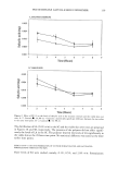
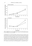
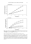
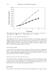
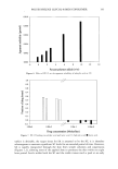
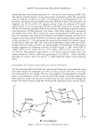
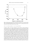
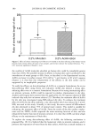
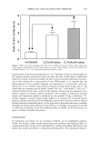
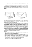
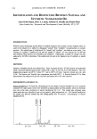
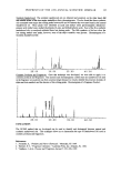
134 JOURNAL OF COSMETIC SCIENCE is 1,800. The mechanism by which the polymer reduced the rates of permeation of actives to the skin is presented. Some factors that could influence the efficacy of the polymer were studied, and the results are reported. EXPERIMENTAL MATERIALS Materials included salicylic acid (SA), dibasic sodium phosphate, monobasic potassium phosphate, sodium chloride, potassium chloride, sodium hydroxide, tetramethylammo- nium hydroxide pentahydrate, acetonitrile HPLC, methanol HPLC grade, scintillation fluid, and glacial acetic acid (Fisher Scientific, Fair Lawn, NJ) lactic acid (LA) (Sigma, Saint Louis, MO) polyethylene glycol-8/SMDI copolymer (Polyolprepolymer-15, Pene- derm Inc., Foster City, CA) hydroxyethyl cellulose (Natrosol 250 HR, Aqualon Com- pany, Wilmington, DE) cellulose acetate dialysis tubing (Spectrum, Houston, TX) poloxamer 105 (Pluronic L-35, BASF, Parsippany, NJ) polyoxyethylene (20) sorbitan monooleate (Tween 80, ICI, Wilmington, DE) diazolidinyl urea, methylparaben, and propylparaben (Sutton Laboratories, Chatham, NJ) [•4C]SA-56.1 mCi/mmol, [•4C]LA- 108.3 mCi/mmol, and [3H]water-108.3 mCi/mmol (NEN Products, Boston, MA) skin-digesting fluid (Solvable, Packard Instrument Company, Inc., Meriden, CT) and aqueous detergent (Palmolive dishwashing liquid, Colgate Palmolive, New York, NY). FORMULATIONS Solutions and suspensions. Buffered aqueous preparations (pH = 2.4) containing various concentrations of SA ranging from 0.1% to 2% w/w were formulated. Formulations containing SA at a concentration higher than 0.2% w/w were suspensions. All formu- lations contained a preservative (0.2% w/w diazolidinyl urea) and a dispersing agent (0.1% w/w polyoxyethylene (20) sorbitan monooelate). When polyethylene glycol-8/ SMDI copolymer (PP-15) was added to the formula, its concentration ranged from 0.3 % to 6% w/w. Water was displaced to accommodate the addition of PP-15. Lactic acid (LA) was formulated in buffered aqueous solutions (pH = 2.0). Its concen- tration in the formulations ranged from 5% to 10% w/w. All formulations contained a preservative (0.2% w/w diazolidinyl urea) and a dispersing agent (0.1% w/w polyoxy- ethylene (20) sorbitan monooleate). In some formulations, 3% w/w PP-15 was added to the formula and water was displaced to accommodate the addition. Aqueous solutions of 0.25 % w/w methylparaben (MP)and 0.004% w/w propylparaben (PP) contained a surface-active agent (0.1% w/w polyoxyethylene (20) sorbitan mono- oleate). When PP-15 was added to the formula, 3% w/w of the water was displaced. Formulations containing 0.5% w/w SA and 5.0% w/w LA were used in most of the experiments unless otherwise stated. IN VITRO PERMEATION Preparation of the skin. Fresh, excised, micro-Yucatan pig skin was supplied by Charles River Laboratories (Wilmington, MA). Upon receipt, the skin was washed gently with

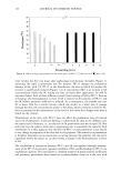
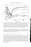
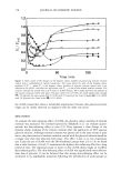
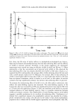
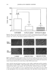
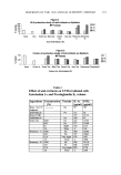
POLYETHYLENE GLYCOL-8/SMDI COPOLYMER 135 1% (v/v) aqueous dishwashing liquid, rinsed with distilled water, and patted dry with a paper towel. A 250-300 pm thick layer of the skin was prepared with a Padgett Electrodermatome (Padgett Dermatome, Division of Kansas City Assemblage Co., Kan- sas City, MO). The dermatomed skin was refrigerated until used. Two hours before each experiment, the skin was placed at room temperature to equilibrate. Circular pieces of the dermatomed skin (about 12 mm in diameter) were cut with a brass punch and placed epidermis-side up on the diffusion cells. Methodology. The skin was cut into discs 12 mm in diameter that were mounted on flow-through (8) diffusion cells (Amie Systems, Riegelsville, PA). The diffusion cells were clamped and the receptor fluid, phosphate-buffered saline (PBS), was pumped through at a rate of 3.57 ml/hour. The membrane was left in place one hour to equili- brate before application of the material to be tested. The cells' temperature was main- tained at 32øC throughout the experiment by means of a water bath/circulator (Haake, Paramus, N J). Fraction collection took place at specified intervals (every hour for SA and every four hours for LA) throughout the experiment by means of a fraction collector (Isco, Inc., Lincoln, NE). Samples were collected directly into scintillation vials. All samples were tested in quadruplicate. Unless otherwise indicated, a 50-pl (low-dose) sample of the formula was dispensed and spread evenly on the skin surface using a micropipette. The cells were left uncovered throughout the experiment. In infinite-dose experiments (500 pl), the cells were covered during the experiment. Skin uptake. The skin was examined for uptake at set intervals: 2, 4, 6, and 8 hours for SA and 4, 8, 12, and 20 hours for LA. Four replicates were tested in each experiment. Before measuring uptake, the skin was washed thoroughly while mounted on the dif- fusion cells. Washing consisted of wiping the skin surface with a 1-cm 2 piece of tissue followed by addition of 100 pl of a 70/30 (v/v) ethyl alcohol/water solution to the surface of the skin and wiping it with another piece of tissue of the same size. Finally, the skin was wiped a third time with a 1-cm 2 piece of tissue, and the three pieces of tissue were set aside for analysis. The skin was then removed from the diffusion cells and tape- stripped. Twenty-seven consecutive tape-strippings were performed on each piece of skin. The first two strippings were added to the three pieces of tissue collected in the wash and were assayed together to determine the amount of active remaining on the skin. The other twenty-five strippings were bundled in groups of five and analyzed for drug content by means of scintillation counter (Beckman Instruments Inc., Fullerton, CA). The twenty-five strippings collected represented the amount of active in the stratum corneum (SC). When the stripping was completed, each piece of skin was digested and assayed for drug content. Digestion was performed by adding 2 ml of skin-digesting fluid and incubating the skin for 48 hours in a 40øC incubator (Precision Scientific Co., Chicago IL). The samples were then removed and brought to room temperature, and 0.1 ml of glacial acetic acid was added to each sample. Drug content was then measured using the scintillation counter. Analysis Radiolabeling. Salicylic acid and LA formulations were spiked with 1 l•l/ml of [•4C]SA 14 . ß - 14 and 4 l•l/ml of [ C]LA, respectively. Each m•crohter of [ C]SA or [•4C]LA contained 0.1 l•Ci (2.2 x 10⁵ DPM). The receptor fluid from all permeation experiments was collected directly into scintillation vials. Ten milliliters of scintillation fluid was added to each vial, and all samples were analyzed in the scintillation counter.
Purchased for the exclusive use of nofirst nolast (unknown)
From: SCC Media Library & Resource Center (library.scconline.org)