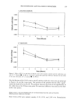
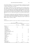
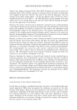
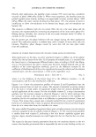
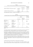
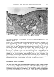
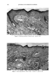
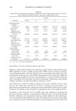
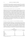
VITAMIN A AND GLYCOLIC ACID FORMULATIONS 167 Table I Mean Karyometric Values of Cells of the Basal Epidermal Layer of Control Guinea Pigs and of Guinea Pigs Topically Treated With Cosmetic Preparations (Mann-Whitney test) Groups Parameter Control T n T m T•v T v Larger diameter (prn) 6.88 9.18' 7.82** 8.68* 8.38* Smaller diameter (pm) 4.79 5.96* 5.52* 5.84* 5.28* Mean geometric diameter (pm) 5.72 7.37* 6.55* 7.09* 6.62* D/d ratio 1.46 1.57' 1.43 ns 1.51 ns 1.61' Volume (pm 3) 101.88 215.71' 151.13' 192.74' 155.83' Area (pm 2) 26.03 43.08* 33.99* 39.91' 34.74* V/A ratio 3.81 4.91' 4.36* 4.73* 4.42* Perimeter (pm) 18.50 24.09* 21.13* 23.06* 21.79' Eccentricity 0.70 0.74 '•s 0.69 '•s 0.71 " 0.75* Form coefficient 0.94 0.93 '•s 0.95 n• 0.94 ns 0.92* Outline index 3.65 3.69** 3.64 n• 3.67 ns 3.71' * p 0.01 **p 0.05 ns, Nonsignificant. Table II Mean Karyometric Values of the Spinous Layer Cells of the Epidermis of Control Guinea Pigs and of Guinea Pigs Topically Treated With Cosmetic Preparations (Mann-Whitney test) Groups Parameter Control T n T m T•v T v Larger diameter (prn) 6.78 9.18* 7.94'* 8.23' 7.21' Smaller diameter (Fro) 4.50 6.10* 5.50* 5.87' 5.68' Mean geometric diameter (Fro) 5.55 7.46* 6.59* 6.93* 6.68* D/d ratio 1.51 1.5 3 ns 1.46 • 1.42'* 1.41 ** Volume (pm 3) 90.95 222.62* 153.13' 178.93' 161.52' Area (pm 2) 24.33 44.05* 34.35* 38.05* 34.46* V/A ratio 3.69 4.97* 4.39* 4.62* 4.45' Perimeter (Fro) 17.81 24.31' 21.333' 22.34* 21.54' Eccentricity 0.73 0.72 ns 0.69** 0.67* 0.66* Form coefficient 0.96 0.94 n• 0.95 • 0.95 n• 0.95 ns Outline index 3.63 3.68 ns 3.65 n• 3.64 n• 3.64 n• *p 0.01 **p 0.05 ns, Nonsignificant. STEREOLOGIC RESULTS The basal layer was stereologically thicker in the areas treated with the formulations, and this was more evident in the presence of glycolic acid. Although thicker, the basal layer presented a smaller number of cells. This increase was compared on the basis of increased cytoplasmic and cellular volumes (Table III). The same was observed with respect to the spinous layer (Table III). When the epidermis was considered as a whole, there was increased thickening, espe- cially after the use of glycolic acid. This increased thickening was also reflected on the decreased surface density. The number of cells per mm 3 was decreased (Table III). The horny layer was not evaluated quantitatively because of the trauma cased by shaving.

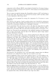
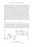
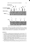
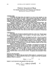
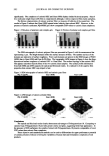
168 JOURNAL OF COSMETIC SCIENCE Table III Mean Values of the Stereologic Parameters of Epidermal Cells of Control Guinea Pigs and of Guinea Pigs Topically Treated With Cosmetic Preparations (Mann-Whitney test) Groups Parameter Control T• T•n T•v T v Germinative layer Cytoplasm volume (m •) 620.33 1010.08' 1016.69' 764.14'* 1323.44' Cell volume 718.94 1220.32' 1164.57' 951.41' 1476.18' N/C ratio 0.16 0.21' 0.15 ns 0.25* 0.12'* Thickness (l•m 3) 7.32 11.51' 11.11' 16.56' 16.26' Numerical density (nø/mm 3) 1438348 825519* 869289* 1059321 * 712823' Spinous layer Cytoplasm volume (l•m •) 1067.43 1901.33' 2144.54' 1473.18' 2552.61' Cell volume (l•m 3) 1157.09 2119.41' 2294.59* 1648.15' 2709.33* N/C ratio 0.08 0.11 * 0.07 ns 0.14 "• 0.06* Thickness (lam 3) 9.36 16.95' 23.71' 29.43* 37.78* Numerical density (nø/mm 3) 876950 476206* 471392' 665843* 374095* Total thickness Surface density (mm2/mm 3 ) 4.76 2.58* 2.38* 1.52' 1.40* Thickness (lam 3) 22.72 34.98** 39.90** 59.59 64.57* Outer layer/basal layer ratio 1.04 1.04 n• 1.01 •s 1.00 • 1.00 '• Numerical density (nø/mm 3) 1011357 487789* 526503' 620644* 401681 * * p 0.01 **p 0.05 ns, Nonsignificant. MECHANISMS OF ACTION OF ALPHA-HYDROXY ACIDS (AHAs) AHAs are widely used in therapy since they have specific effects on the skin structures. When applied to the skin in high concentrations, AHAs cause the release of keratino- cytes and epidermolysis, and when applied at low concentrations they reduce the cohe- sion between corneocytes and cause evident desquamation of the horny layer. AHAs are involved in many metabolic processes: they participate in essential cellular mechanisms such as the Krebs cycle, glycolysis, and serine biosynthesis. Furthermore, they promote collagen maturation and glycosaminoglycan formation (10,11). The reduction in corneocyte cohesion caused by the action of AHAs occurs at lower levels in the horny layer, and this fact involves a dynamic and active process in a particular step of keratinization. Among the various possibilities, there may be a modi- fication of ion binding or the dissolution of desmosomes due to the reduced pH (11). According to Van Scott and Yu (12), AHAs reduce the cohesion of corneocytes by affecting ion bonds, as follows: 1) by hydration of the horny layer, increasing the distance between corneocytes and consequently reducing cellular cohesion. In our material gly- colic acid provoked a deep hydration not only in the horny layer, but also in the spinous one, resulting in greatly thickened epithelium and wide separation between cells 2) by
Purchased for the exclusive use of nofirst nolast (unknown)
From: SCC Media Library & Resource Center (library.scconline.org)