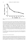
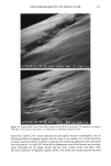
74 JOURNAL OF COSMETIC SCIENCE information could be obtained by studying intact individual hair fibers, but the small diameter of the hair fibers makes such measurements difficult (100 pm). The problems associated with the small sample dimensions have been overcome (7,8) by combining the chemical specificity afforded by infrared spectroscopy with the lateral resolution of IMS. Indeed, using a double-aperture experimental arrangement to reduce spurious signals from neighboring areas (9), regions down to =30 microns in size have been analyzed (10). Though individual hair shafts are small, their thickness still exceeds the optical penetration depth at wavelengths where absorption is quite strong. By means of IMS, flattened hair shafts treated with oxidizing agents have been examined at different distances from the root to the tip (6). Using a diamond "squeeze-cell" to effectively thin the hair, characterization by IMS of hair samples from the anagen to the telogen phases has also been made by probing the fiber from the bulb to the shaft (11) while the sensitivity of cortical hair cells in contact with potassium hydroxide in 1-butanol solutions has been tested using IMS (12). IMS has also been employed to examine the inner part of a microtomed hair to determine drug ingestion (13-15), and absorbance contour linear maps have been obtained. Nevertheless, IMS experiments using a thermal source (globar) are limited, by the brightness of the source, to a lateral resolution above 24 x 24 pm (5,10). Replacing the traditional thermal source of a conventional IR microscope with a high-brightness synchrotron source allows substantial improvements of both signal-to-noise ratio and lateral resolution of IMS measurements (16,17). Thus, experiments can be carried out in diffraction-limited conditions, and an image of a hair sample can be obtained with a lateral resolution of few microns. IMS with synchrotron radiation was recently performed to study the localization of drug metabolites within longitudinally microtomed sections of human hair (15). In this investigation, a synchrotron IMS study of hair shaft samples was performed. We present for the first time--detailed, high contrast, chemical images of transverse hair cross sections. In particular, the cortex, cuticle, and medulla are differentiated. Results are compared with those obtained using a standard thermal source. Bleached hair was also imaged chemically with a lateral resolution of a few microns. MATERIALS AND METHODS SAMPLE PREPARATION Untreated, dark homogeneous human hair fibers of European origin were used. Tresses approximately 15 cm in length were first separated into 2-g pieces. A bleaching cream was prepared by blending one volume of commercial bleaching powder (Platifiz © from l'Oreal Inc.) with two volumes of hydrogen peroxide solution (40 volume). To complete the bleaching process, bleaching cream was uniformly applied on dry hairpieces (10 g per 2-g piece). Samples were then wrapped in an aluminum sheet and left at ambient temperature for one hour. The hair samples were then washed with water and retreated with the bleaching cream for another hour. Finally, the hair samples were water rinsed and allowed to dry at ambient temperature. SAMPLING FOR IMS EXPERIMENTS For the IMS experiments, the hair fibers were first cut into 1-cm lengths starting

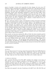
CHEMICAL IMAGING OF HAIR BY IMS 75 approximately 10 cm from the root. Then these were embedded into epoxy resin and sliced with a microtome to yield cross sections 6-pm thick. The polymer was chosen to have spectral features that overlapped minimally with those of the hair sample. INFRARED MICROSPECTROSCOPY EXPERIMENTS The infrared data were obtained with a commercial IR microspectrometer (Spectra-Tech "Irps TM) installed at beamline U4IR (16,17) at the National Synchrotron Light Source, Brookhaven National Laboratory (USA). The high-brightness infrared synchrotron radiation (IRSR) was extracted from the elec- tron storage ring using special high-aperture f/10 optics, and a collimated beam of 2-cm diameter was fed into the IrpS microspectrometer system. A schematic of the Irps microspectrometer is shown in Figure 1. The IrpS system consists of an integrated FTIR spectrometer and a microscope optical module. The spectrometer is equipped with a rapid-scan Michelson interferometer with a Germa- nium-coated KBr beam splitter and a mercury cadmium telluride (MCT) infrared de- tector. A 32X, 0.65 numerical aperture (NA) objective and 10X, 0.71 NA condenser were in a confocal arrangement for the transmission measurements. For our measure- ments, apertures were placed both before and after the sample to confine the infrared beam to a particular region of interest in the specimen and to provide maximum rejection of stray light, as shown by Sommer and Katon (9). The aperture sizes quoted in this paper are those defined at the sample, taking account of the appropriate demag- nification of objective and condenser. Spectra were collected between 4000 and 900 cm- • at a resolution of 4 ø cm-•. Infrared Sample on stage X-Y Lower a• Illuminator : sour•3e Upper aperture '•" "• ...... .•.•:" '•"•'•••••iii • .::. :' •7 ••e•spl Detor Figure 1. Schematic diagram of the I•s scanning infrared microsp•trometer.
Purchased for the exclusive use of nofirst nolast (unknown)
From: SCC Media Library & Resource Center (library.scconline.org)