ANTIMELANOGENIC EFFECTS OF HMF 119 at 4°C to obtain clear cell lysates. Western blotting was performed as previously de- scribed (16). Aliquots of the cell lysates (20 µg protein each), diluted in a Laemmli buffer containing 2% SDS and 1.2% dithiothreitol, were heated at 95°C for five minutes to denature the proteins. The proteins were separated by electrophoresis on a 10% SDS- polyacrylamide gel at 200 V for 1.5 hours and transferred to a nitrocellulose membrane. The membrane was incubated with a primary antibody overnight at 4°C, and then with a secondary antibody conjugated with alkaline phosphatase for one hour at room tem- perature. The bands were detected using a chemiluminescence kit (Pierce, Cheshire, UK) according to the manufacturer's instruction, and were subjected to densitometry analy- sis. REVERSE TRANSCRIPTASE-POLYMERASE CHAIN REACTION (RT-PCR) ANALYSIS PCR primer sets specific for mouse tyrosinase and D-glyceraldehyde-3-phosphate de- hydrogenase (GAPDH) were designed using Primer3 software (Whitehead Institute/MT Center for Genome Research) on the basis of sequences deposited in the NCBI GenBank database. The sequences of the primers used in this study were: tyrosinase (GeneBank accession number, NM011661) 5'-ATG GGT CAA CAC CCA TGT TT-3' (sense) and 5'-GGC AAA TCC TTC CAG TGT GT-3' (antisense) and GAPDH (GeneBank ac- cession number, NM008084), 5'-TGT TCC TAC CCC CAA TGT GT-3' (sense) and 5'-TGT GAG GGA GAT GCT CAG TG-3' (antisense). Total cellular RNA was isolated using TRIZOL® Reagent (Invitrogen, Carlsbad, CA) as per the manufacturer's instructions. RT-PCR was performed using GeneAmp® PCR system 9700 (Applied Biosystems, Foster City, CA) in a reaction mixture (20 µl) containing Maxime RT-PCR PreMix (iNtRON Biotechnology, Seongnam, Korea), 500 ng RNA, and 20 pmole of gene-specific primer sets. A reverse transcription reaction for the first strand cDNA synthesis was performed at 45 ° C for 30 minutes, followed by heat inactivation of reverse transcriptase at 95°C for five minutes. A PCR reaction was carried out for 32 cycles of 45 seconds at 95°C, 45 seconds at 55°C, and 60 seconds at 72°C, with a final extension step of five minutes at 72°C. Preliminary PCR runs ascertained that the reactions were in the exponential phase suitable for the quantitative analysis of tyrosinase and GAPDH mRNA. Amplification products were electrophoresed in a 1.2% agarose gel with a 100 bp DNA ladder as a size marker. The gel was ethidium bromide-stained and the band intensities were quantified using a Gel Doc system (BioRad, Hercules, CA). ASSAY OF TYROSINASE ACTIVITY IN VITRO AND IN THE CELLS The in vitro tyrosinase activity was determined by measuring the oxidation of DOPA to DOPA chrome (17). Briefly, the reaction mixture (200 µl) containing cell lysates as a tyrosinase enzyme source (20 µg protein), test materials at different concentrations (0 ~ 1600 µM), and 1.0 mM DOPA as a tyrosinase substrate in 0.1 M sodium phosphate buffer (pH 6.8) was incubated at 3 7°C for 60 minutes. The DOPA chrome formation was determined at 490 nm. To assess the in situ tyrosinase activity, the cells were supplied with 1.0 mM DOPA and incubated for 48 hours. The cell pigmentation was monitored under a microscope. STATISTICAL ANALYSIS Statistical analysis was performed by t-test. The p 0.05, based on at least three or more independent experiments, was considered to be statistically significant.

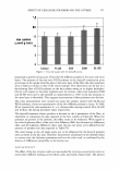
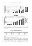
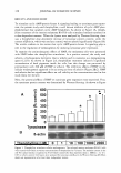
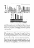
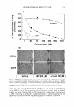
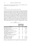
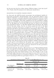
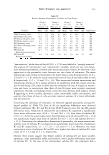
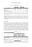
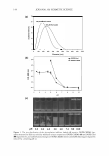
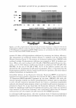
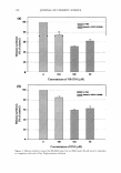
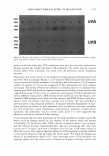
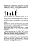
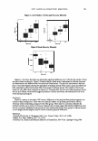
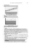
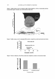
120 JOURNAL OF COSMETIC SCIENCE RESULTS AND DISCUSSION To stimulate cyclic AMP/protein kinase A signaling leading to tyrosinase gene expres- sion, the present study used theophylline, a well known inhibitor of cyclic AMP phos- phodiesterase that catalyzes cyclic AMP breakdown. As shown in Figure lA, theoph- ylline treatment of the murine melanoma B16F10 cells stimulated melanin synthesis in a dose-dependent manner. When the lysates were analyzed by Western blotting, there was a theophylline dose-dependent increase of tyrosinase protein content, while the content of �-actin, which was used as a control, was not altered significantly (Figure 1B). The results conform to the notion that cyclic AMP/protein kinase A signaling plays a role in the regulation of melanogenesis by inducing tyrosinase gene expression. To examine the antimelanogenic effects of HMF, the melanoma cells were pretreated with HMF before the theophylline stimulation. As a positive control, the study used arbutin, a hydroquinone derivative that is widely used in cosmetics as a depigmenting agent (12,18). As shown in Figure 2A, theophylline treatment induced a significant accumulation of dark pigments inside the cells, but this change was prevented by pretreatment with 200 µM of HMF or arbutin. The inhibitory effects of HMF on the cellular melanogenesis appeared to be as strong as those of arbutin (Figure 2B,C). HMF and arbutin had no significant effects on cell viability at the concentrations used in this study (data not shown). Next, the potential effects of HMF on tyrosinase gene expression were examined. First, the tyrosinase protein content was determined by Western blotting. As shown in Figure .-. 600 - A 0 500 .... C 0 400 u � 300 Cl ._. C 200 C ca 100 - CD ::il 0 Tyrosinase B B-actin I Theophylline (µM) I 0 * * * 1 1 1 o 11 oo 11 ooo 1 2000 1 Figure 1. Theophylline stimulates cellular melanogenesis. The cultured murine melanoma B16F10 cells were treated with vehicle or various concentrations of theophylline for 72 hours. The melanin content in the cell-free culture media is expressed as % of vehicle control (A). Data represent mean ± SEM (n = 3). *p 0.05 vs vehicle control. Cell lysates were analyzed for tyrosinase and (3-actin by Western blotting (B). Blots shown are representative of at least three independent studies.
Purchased for the exclusive use of nofirst nolast (unknown)
From: SCC Media Library & Resource Center (library.scconline.org)