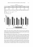
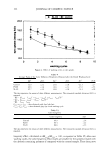
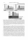
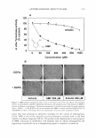
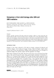
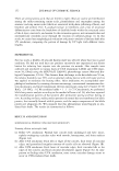
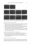
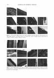
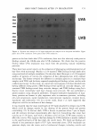
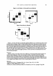
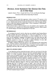
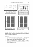
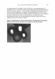
156 JOURNAL OF COSMETIC SCIENCE cortex together, may appear greater after UV A irradiation. On the other hand, UVB causes severe morphological damage, confined to the hair cuticles because of its restricted depth of penetration. CONCLUSIONS In summary, our morphological study shows relatively more destructive cuticular changes after UVB irradiation than after UV A, while disruptions of the intercellular lipid layer show similar results between UVA and UVB irradiation. However, in labile protein analysis, damaged labile hair proteins are much more observed after UVA irradiation than after UVB irradiation. Because high doses of UV light were irradiated to observe patterns of damage in this report, we'd like to plan to observe chronically photodamaged hair shafts as similar as possible to those in daily life. Based on the principal findings here, we hope it will be useful to study the photoaging of hair and photoprotective methods in hair. Other biochemical methods, including amino acid analysis and lipid analysis, would be helpful to understand the process of UV-light- induced damage to hair. ACKNOWLEDGMENTS This research was supported by Health Fellowship Foundation grants in Korea. REFERENCES (1) "Oxidation of Hair Proteins and the Cell Membrane Complex by Sun and Ultraviolet Light," in Chemical and Physical Behavior of Human Hair, 4th ed., Clarence R. Robbins, Ed. (Springer-Verlag, New York, 2002), pp. 163-171. (2) R. Beyak, G. S. Kass, and C. F. Meyer, Elasticity and tensile properties of human hair. II. Light radiation effects,]. Soc. Cosmet. Chem., 22, 667-678 (1971). (3) R. Arnaud, G. Perbet, A. Deflandre, and G. Lang, ESR study of hair and melanin-keratin mixtures- The effects of temperature and light, Int. J. Cosmet. Sci., 6, 71-83 (1984). (4) E. Hoting, M. Zimmermann, and H. Hocker, Photochemical alterations in human hair. Part II: Analysis of melanins,]. Soc. Cosmet. Chem., 46, 181-190 (1995). (5) H. F. Launer, Effect of light upon wool. Part IV. Bleaching and yellowing by sunlight, Textile. Res.]., 35, 395-400 (1965). (6) A. S. Inglis and F. G. Lennox, Studies in wool yellowing. Part IV. Changes in amino acid composition due to irradiation, Textile. Res.]., 33, 431-435 (1963). (7) W. S. Lee, T. H. Oh, S. H. Chun, S. Y. Jeon, E. Y. Lee, S. Lee, et al, Integral lipid in human hair follicle,]. Invest. Dermatol. Symp. Proc., 10, 234-237 (2005). (8) T. Inoue, M. Ito, and K. Kizawa, Labile proteins accumulated in damaged hair upon permanent waving and bleaching treatments,]. Cosmet. Sci., 53, 337-344 (2002). (9) D. Braida, C. Dubief and G. Lang, Photoageing of hair fiber and photoprotection, Skin Pharmacol., 7, 73-77 (1994). (10) V. Bousquet, D. Black, C. Liviero, J.M. Laquarde, and Y. Gall, Analysis of cuticle relief for hair photoprotection evaluation: Validation study, Curr. Prob. Dermatol., 26, 196-202 (1998). (11) A. Hershko and A. Ciechanover, The ubiquitin system, Annu. Rev. Biochem., 67, 425-479 (1998). (12) A. C. Santos Nogueira, and I. Joekes, Hair color changes and protein damage caused by ultraviolet radiation,]. Photochem. Photobiol. B., 74, 109-117 (2004).

]. Cosmet. Sci.) 59, 157-158 (March/April 2008) Abstracts Journal of the Society of Cosmetic Chemists Japan Vol. 41, No. 4, 2007* An Application Study of Culture Supernatant of a Construction of a Three-Dimensional Human Skin Model Filamentous Fungus Isolated from Marine Environment as Consisting of Keratinocytes, Dendritic Cells and an Ingredient for Whitening Cosmetics Fibroblasts and Application of This Model for Alternative Katsuhisa Yamada*, Chiaki Imada**, Takahiro Tsuchiya***, Katsushiro Miyamoto***, Hiroshi Tsujibo***, Takeshi Kobayashi**, Naoko Hamada-Sato** *DHC Corporation, 2-7-1, Mi-nami-azabu, Minato-ku, Tokyo 106-8571, Japan, **Tokyo University of Marine Science and Technology, 4-5-7, Konan, Minato-ku, Tokyo 108-8477, Japan, ***Osaka University of Pharmaceutical Science, 4-20-1, Nasahara, Takatsuki 569-1094, Japan The whitening effect of a culture supernatant from a fungus ofTrichoderma sp. isolated from a marine environment was investigated. The supernatant had tyrosinase inhibitory activity in vitro. From the time course study of the production of tyrosinase inhibitor, maximum production was obtained after incubation for 3 to 4 days. The culture supernatant was subjected to a high performance liquid chromatography in order to investigate the whitening compounds in it. At least three compounds were found in the supernatant having inhibitory activity in vitro. The evaluation by a B 16 cell in all fractions revealed that three active fractions exited. These fractions did not always coincide with the fraction of inhibitory activity in vtro. The fraction with inhibitory activity both in vitro and against B16 cell showed the remarkable inhibitory effect of melanogenesis. Thus, the culture supernatant contained various components having whitening effects, suggesting that it is possible to apply it to new whitening cosmetics. Animal Testing oflmmune-Sensitizing Compounds Tadashi Uchino, Yoshiaki lkarashi, Hiroshi Tokunaga National Institute of Health Sciences, 1-18-1, Kamiyoga, Setagaya-ku, Tokyo 158-8501, Japan In order to establish in vitro evaluation of the sensitization of human skin, we attempted to make a three-dimensional human skin model consisting of three different cells, dendritic cells, keratinocytes and fibroblasts. The viability of the cells in the human skin model was observed after staining with hematoxylin and eosin. After 11-14-day incubation (horny layer was initially observed), the three- dimensional human skin model was used for experiments. Due to 2,4-dinitrochlorobenzene (DNCB) under a non- cytotoxic dose, the keratinocytes and dendritic cells in the human skin model significantly induced IL-4 release into the incubating medium and dendritic cells induced CD86 expression. On the other hand, with sodium dodecyl sulfate (SOS non-sensitizer), the keratinocytes and dendritic cells did not significantly induce IL-4 release and the dendritic cells did not induce CD86 expression. The results suggested that this three-0imensional human skin model with dendritic eel Is could be applied as an alternative to animal testing ofimmune-sensitizing compounds. * These abstracts appear as they were originally published. They have not been edited by the Journal of Cosmetic Science. 157
Purchased for the exclusive use of nofirst nolast (unknown)
From: SCC Media Library & Resource Center (library.scconline.org)