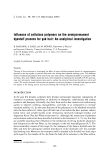
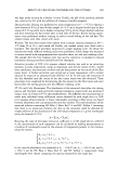
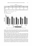
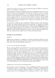
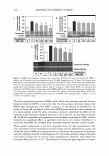
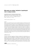
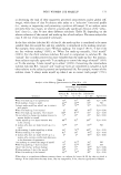
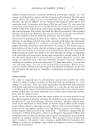
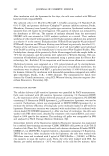
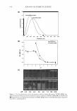
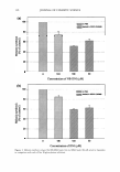
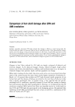
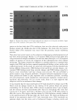
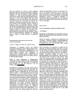
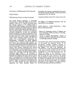
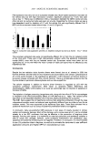
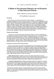
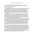
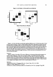
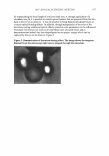
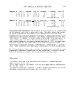
144 JOURNAL OF COSMETIC SCIENCE (a) 30 25 100 µM NB-DNJ loaded i 20 15 10 en 5 o o 100 200 300 400 500 600 700 800 900 1000 Diameter [nm] (b) ....._oOPE:Chol 0.8 __.,_ DOPE:CHEMS o.e. e 0.4 0.2 0.1) -0.2 ... ? 4 �. e 7 8 9 10 pH (c) pH 2.5 3.0 4.5 5.0 6.0 7.4 8.0 10.0 Figure 1. The size distribution of the glycosylation inhibitor-loaded pH-sensitive DOPE:CHEMS lipo- somes measured by DLS (a) turbidity measured using a suspension of DOPE:CHEMS (.&) and DOPE:Chol (e) liposomes (b) and conformational changes ofDOPE:CHEMS (lower) and DOPE:Chol (upper) liposomes induced by a lowering pH (c).

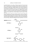
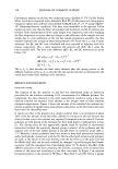
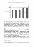
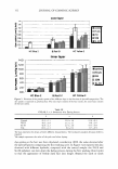
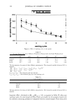
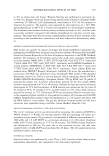
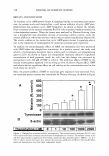
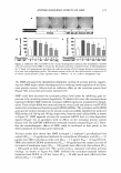
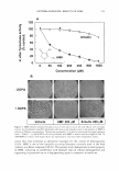
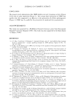
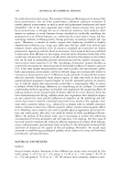
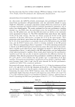
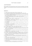
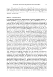
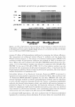
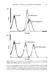
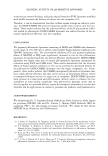
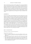
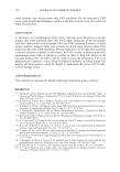
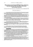
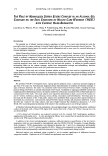
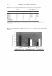
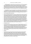
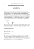
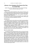
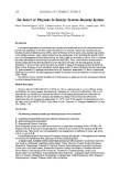
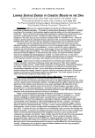
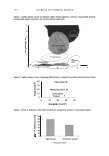
DELIVERY ACTIVITY OF pH-SENSITIVE LIPOSOMES (a) 80 kDa 70 60 µM NB-DNJ 0 (b) 0 loaded in DOPE:CHEMS 200 50 200 100 loaded in DOPE:CHEMS 100 100 50 µMNB-DNJ 100 50 5 loaded in DOPE:CHEMS 100 50 µMDNJ 5 145 Figure 2. GI effects of glycosylation inhibitor-loaded pH-sensitive liposomes in comparison with the free N-glycosylation inhibitors evaluated by Western blotting: GI effects depending on various concentrations of NB-DNJ loaded or not loaded in DOPE:CHEMS liposomes (a) and enhanced GI effects by NB-DNJ- loaded or DNJ-loaded DOPE:CHEMS liposomes (b). increase GI effects of N-glycosylation inhibitors by increasing the delivery efficiency. The measurement of synthesized melanin (Figure 3) correlates with the results from Western blotting (Figure 2). The amount of synthesized melanin from HM3KO cells incubated without N-glycosylation inhibitors was assumed as 100% of melanin syn- thesis. When cells were incubated with 100 µM of NB-DNJ-loaded pH-sensitive lipo- somes, 22.8 % of the melanin synthesis was reduced in comparison with 100 µM of free NB-DNJ. In the case of 100 µM of DNJ, 26.1 % of the melanin synthesis was also decreased by the incorporation of pH-sensitive liposomes. Thus, it was found that the N-glycosylation inhibitor, which is loaded in pH-sensitive liposomes, efficiently reduced melanin biosynthesis in mammalian cells. Intracellular delivery of two fluorescent molecules, fluorescein-DHPE incorporated in liposomes and calcein loaded in liposomes was evaluated by FACS. After incubation with 1 % fluorescein-DHPE-embedded liposomes for one hour, HM3KO cells with pH- sensitive liposomes resulted in a relative strong fluorescent intensity value in comparison to cells with pH-insensitive liposomes (Figure 4a). Calcein, a membrane-impermeable molecule, also showed higher intensity when it was loaded in pH-sensitive liposomes, as presented in Figure 46. It was found that the fluorescent intensities of fluorescein, a marker for components of pH-sensitive liposomes and calcein, and a tracer as a delivered cargo material in pH-sensitive liposomes, were significantly higher than the observed intensity from the cells treated with pH-insensitive PC:CHEMS liposomes. In additional CLSM experiments, cells were incubated with 1 % fluorescein-embedded liposomes (L) containing dextran-rhodamine B to better define the intracellular deliv-
Purchased for the exclusive use of nofirst nolast (unknown)
From: SCC Media Library & Resource Center (library.scconline.org)