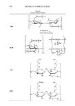
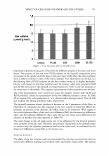
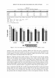
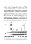
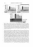
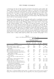
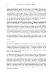
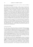
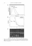
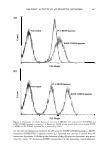
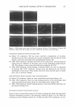
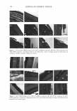
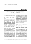
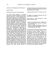
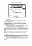
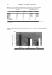
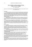
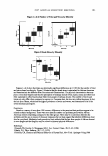
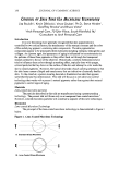
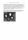
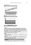
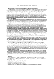
168 JOURNAL OF COSMETIC SCIENCE KEVNOTE AWARD LECTURE SPONSORED BV RUGER CHEMICAL CORPORATION NON-SUNSCREEN PHOTO PROTECTION: WHERE ARE WE TODAV? THE SKIN IMMUNE PROTECTION Lieve Declercq, Ph.D., Daniel Maes, Ph.D., Thomas Mammone, Ph.D. and Mary Matsui, Ph.D. Estee Lauder Companies, Inc. Today's scientific literature abound with solid evidence that the various parts of the UV spectrum do generate significant damage in the skin, and that the necessity to provide a broad spectrum protection from UV exposure is not just an option, but has become an absolute necessity. Despite the significant improvement in sunscreen technologies observed recently, based mostly on the stabilization of the UV absorbers during sun exposure, it remains that sunscreens have not been capable so far to provide a complete protection from al I the consequences related from the exposure to both UVB and UVA rays. Clearly, the damages caused by the generation of free radicals, and the resulting oxidation of lipids proteins and even DNA cannot be totally prevented even by the best combination of the existing sunscreens technology. One of the most deleterious consequences of UV exposure is the suppression of the cutaneous immune response, as it is clearly associated with skin cancer formation. The process by which UV suppress immunity involves a complex combination of pathways that includes oxidation , inflammation, damage to DNA, leading to the depletion and alteration of antigen presenting cells. DNA repair enzymes such as T4 endonucleases as well as fragments of RNA have been shown to stimulate the repair of UV-induced DNA lesions, as well as prevent the UV-induced up regulation of immuno-suppressive cytokines such as IL-10 in human skin. Similarly, the free radical-induced oxidative damages have been shown to affect significantly the antigen presentation capacity of the Langerhans cells leading to a significant disruption of the skin immune defense activity. In order to develop a protection technology aimed at preventing the UV-induced immuno suppression, we tested both ex vivo and in vivo, the efficacy of either anti-oxidants or DNA repair technology in maintaining the skin's immune response even after exposure to the full UV light spectra. Ex vivo experiments Experiments were first carried out ex vivo on skin explants maintained in survival. Exposure to solar simulated light caused a dose-dependent increase in sun burn cells, coinciding with a reduction in the number and dendricity of LC. The LC depletion seemed to occur at doses lower than those required to induce formation of sunburn cells. This model was used to evaluate the protective benefits provided by a formulation containing white tea, which was shown previously to have strong antioxidant activity. Skin samples (Non-treated and vehicle treated, as well as skin samples treated with the white tea containing formulation) coming from the same donor were irradiated with increasing doses of UV light (0.45 to 3.56 J/cm2) using an Oriel Solar simulator, corresponding to an estimated 0.25 to 3.0 MED. Treatment with the lotion was performed 4 hours before exposure to UV light. The skin samples were processed 24 hours after exposure for evaluation of the Langerhans cells using immunolabeling with CD I a antibody. The results (Fig. I) clearly demonstrate the protective benefits provided by the white tea containing formulation in preventing the UV-induced depletion of Langerhans cells in the epidermis. The tissue treated with the white tea containing formulation showed no decrease in the number of LC per mm epidermis, up to a dose of 3.56 J/cm2 in the current experimental conditions, which is expected to be approximately equivalent to 2 MED for this phototype. The vehicle formulation did not offer any protection against UV-induced LC migration.

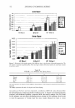
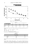
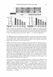
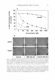
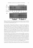
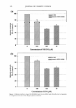
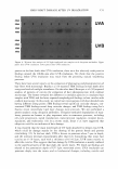
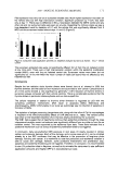
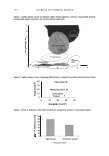
2007 ANNUAL SCIENTIFIC MEETING 18 16 E E 14 ... 12 0 .. .! 10 I C 8 6 0 In vivo experiments: Dose-dependent effect of SSR on Langerhans cells In ex vivo skin treated with white tea ■\8hicle • \\hite tea 2 3 4 SSR do• (J/cm2) Figure 1 This study aimed to compare the effect of topical formulations containing T4 Endonuclease liposomes, RNA fragments, or a lotion containing white tea on the prevention of UV-induced contact hypersensitivity suppression in humans. 169 100 subjects ofFitspatrick Skin type I to III and MED ranging between 20-50 mj/cm2 were randomized into 5 groups which received no treatment, vehicle, or one of the above preparations. Products were selfapplied on gluteal skin once daily for 6 days. Halfofthe subjects per group received solar simulated radiation at 0. 75 MED, over the treatment site on day 3 of product application. DNCB sensitization followed 3 days after irradiation. The contact hypersensitivity response was measured 2 weeks after sensitization by the increase of skin fold thickness over 5 elicitations on the arm. Effect of a 0.2% white tea preparation on the UV-induced immunosuppression: Results clearly demonstrate that the treatment with the white tea containing product resulted in a significant protection against the UV-induced langerhans cells depletion (22% reduction in CD la positive cells after UV exposure and treatment with the white tea, vs. 57% reduction after UV exposure alone), and as a result prevented most of the UV-induced Contact Hypersensitivity suppression (27% reduction in CHS after UV exposure and treatment with the white tea, vs. 53% reduction in CHS after exposure to UV light alone). Similarly, the experiments conducted with a preparation containing either 1 % micrococcus lysate (T4 endonuclease), or 1% RNA fragments, both provided protection from UV-induced Langerhans cell depletion as well as UV-induced Contact Hypersensitivity Suppression (63% reduction of CHS after exposure to UV alone vs. 18% reduction after UV and treatment with the Micrococus lysate). Conclusions: The combination of the Ex vivo and In vivo experiments clearly demonstrate the effect of protecting the skin celJs from oxidative damage as well as increasing the natural DNA repair process results in the prevention the UV-induced immuno suppression. This effect seems to originate from the inhibition of the UV-induced migration of the Langerhans cells away from the skin. Clearly, the combination of this technology with sunscreens enhances significantly the protective benefits provided by the latter. These technologies altogether will allow for the development of products which will provide a far broader protection than what is obtained actually with topical sunscreens formulations.
Purchased for the exclusive use of nofirst nolast (unknown)
From: SCC Media Library & Resource Center (library.scconline.org)