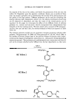
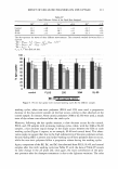
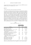
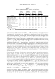
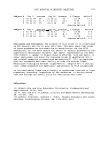
140 JOURNAL OF COSMETIC SCIENCE and their cargo materials may be degraded by various hydrolases and peptidases in the lysosomes. The pH-sensitive liposomes have been designed to circumvent this lysosomal degradation by releasing their cargo contents prior to reaching the lysosomes or partly into the cytosol, where they can then diffuse to target sites (1,2). An amphiphilic stabilizer such as CHEMS incorporated in phosphatidylethanolamine (PE)-based lipo- somes is protonated, and their conformation is changed by acidic environments when they are delivered into endosomes (3,4). N-glycosylation inhibitors such as DNJ and NB-DNJ have an cx-glucosidase-inhibiting effect, which is a useful property for enhanc- ing pigment lightening on mammalian skin by interfering with the maturation of tyrosinase (5,6). However, it is difficult for these inhibitors to translocate through skin and even cellular membranes due to their hydrophilicity (7 ,8). Therefore, DNJ should be delivered across the skin and into the cytoplasmic active site by an efficient delivery carrier to facilitate biological activity for pigment-lightening effects with a minimum concentration in vitro and in vivo. In this study, we attempt to evaluate the N- glycosylation-inhibiting (GI) effects of the pH-sensitive liposomes containing N- glycosylation inhibitors on human melanoma cells, HM3KO, to see the possibility of cosmetic application as a delivery carrier of depigmentation active molecules. Further- more, the in vitro delivery efficiency of pH-sensitive liposomes was examined to confirm the location of the intracellular-delivered pH-sensitive liposomes. EXPERIMENTAL PREP ARA TI ON OF LIPOSOMES Liposomes were prepared according to lipid hydration methods. Molar ratios of lipid components of CHEMS were fixed at 3:2. Compositions of the prepared liposomes are listed in Table I. Briefly, a mixture of lipids in chloroform/methanol (95:5) was dried using a rotary evaporator under reduced pressure. Dried lipids were hydrated with PBS containing N-glycosylation inhibitors to be loaded. Hydrated lipid films were sonicated using a bath-type sonicator. The size distribution of the resulting liposomes was mea- sured by dynamic light scattering (DLS) with a vertically polarized He-Ne laser (Zeta- sizer 3000HS, Malvern, UK). Turbidity was observed by the absorbence of a liposomal Number Table I Formulations of Prepared Liposomes Lipid composition aDOPE:6CHEMS CPC:CHEMS DOPE:dfluorescein-DHPE:CHEMS PC:fluorescein-DHPE:CHEMS DOPE:fluorescein-DHPE:PEG-5 rapeseed sterol DOPE:cholesterol a 1, 2-Dioleoyl-sn-glycero-3-phosphoethanolamine. 6 Cholesteryl hemisuccinate. c L-a-Phosphatidylcholine. d N-(fl uorescein-5-thiocarbamoyl)-1,2-dihexadecanoy1-sn-glycero-3-phosphoethanolamine.

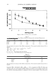
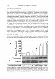
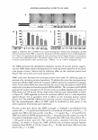
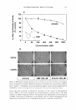
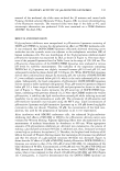
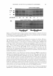
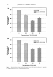
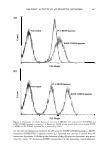
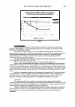
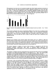
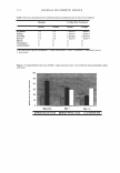
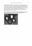
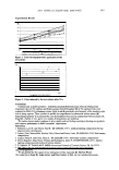
DELIVERY ACTIVITY OF pH-SENSITIVE LIPOSOMES 141 suspension at 500 nm using a Cary 3E UV-VIS spectrophotometer (Varian, Victoria, Australia) to evaluate the pH-sensitivity of the prepared liposomes (15). To quantify intracellular delivery of pH-sensitive liposomes by FACS measurement, either a component of the lipid bilayer or a loading material was marked with fluores- cence. To label the lipid bilayer, DOPE, CHEMS and 1 % N-(fluorescein-5-thiocar- bamoyl)-1,2-dihexadecanoyl-sn-glycero-3-phosphoethanolamine (fluorescein-DHPE) (w/w) (Ex/Em = 495/519 nm Molecular Probes Inc., OR) was dissolved in chloroform/ methanol, and then dried and hydrated (L3 liposome). Fluorescein-encapsulated pH- sensitive liposomes were made by hydration of DOPE:CHEMS (3:2) film with 75 µM calcein (Ex/Em = 494/517) in PBS. 1-cx-Phosphatidylcholine (PC):CHEMS (3:2) was used as a control. After hydration, the procedure of liposome preparation was as previ- ously described. For the CLSM study, a dried film of DOPE:CHEMS (3:2) containing 1 % fluorescein- DHPE (Ex/Em= 495/519 nm) (L3 liposome) was hydrated with PBS containing 2.5 µM dextran-rhodamine B (10,000 mw (Ex/Em = 572/589 nm Molecular Probes Inc., OR). Therefore, the lipid bilayer and loading materials were labeled simultaneously to trace the cellular uptake of the pH-sensitive liposomes. PEG-5 rapeseed sterol (Cognis GmbH, Diisseldorf, Germany) was used instead of CHEMS as an amphiphilic stabilizer for a control L5 liposome. Unencapsulated fluorescence was separated from the liposomes by using MicroSpin™ G-25 columns (Amersham Biosciences Corp., NJ). CELL CULTURE HM3KO, a pigmented melanoma cell line (9), was cultured with minimum essential medium (MEM) supplemented with 1 % (v/v) antibiotics (streptomycin, 10,000 µg/ml penicillin, 10,000 IU/ml) and 10% (v/v) FBS (Gibco BRL, MD) at 37°C in a humidified atmosphere containing 5% CO2 . The cells were subcultured every five days. For the cytotoxicity assay, the suspension of the cells was poured into a 96-well flat-bottomed plate. After adhesion to the plate, cells were incubated with a 2.8-360 µM lipid concentration of liposomes in 100 µl of culture media for an additional 48 hr, and the cytotoxicity was then evaluated by 3-(4,5-dimethylthiazol-2-yl)-2,5-diphenyltetrazo- lium bromide (MTT) assay (10). Briefly, MTT was dissolved in phosphate buffer at 5 mg/ml and filtered for sterilization. Ten microliters of the MTT solution was added to the medium, resulting in a final concentration at 500 µg/ml, and then the cells were incubated for 4 h at 3 7°C. The purple formazan product was dissolved by 100 µ1/well of 40 mN HCl in isopropyl alcohol. Absorbence was measured at 550 nm in a microplate reader (Elx800, Bio-TEK Instr. Inc.). PIGMENT LIGHTENING Pre-cultured HM3KO cells were incubated with culture media containing 1 to 200 µM of NB-DNJ or DNJ-loaded DOPE:CHEMS liposomes (L1 ), for an additional five days. Concentration of the added liposomes was determined by the MIT assay evaluating the viability of the cells. The medium containing liposomes was renewed every two days.
Purchased for the exclusive use of nofirst nolast (unknown)
From: SCC Media Library & Resource Center (library.scconline.org)