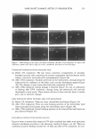
142 JOURNAL OF COSMETIC SCIENCE After incubation with the liposomes for five days, the cells were washed with PBS and harvested with trypsin/EDTA. The collected cells in 0.1 M ofTris-HCl (pH 7.2) buffer containing 1 % Nonidet P-40, 0.01 % SDS, and protease inhibitors (Complete™ protease inhibitor mixture Roche, Mannheim, Germany) were lysated by probe-type sonication. Synthesized melanin was separated from cell lysates by centrifugation. The quantity of melanin was measured by the absorbance at 490 nm. The amount of melanin obtained from the non-treated HM3KO cells was assumed as 100% melanin synthesis as a control. Thus, melanin synthesis of cells treated with liposomes or free N-glycosylation inhibitors was presented as a relative % to the control. Endoglycosidase H (EndoH) and peptide-N-glycosidase F (PNGaseF) digestion and Western blotting were followed as previously described (11). Proteins of the cell lysates (10 µg of protein in 5 µl of cell lysis buffer) were hydrolyzed with EndoH according to the manufacturer's instructions (New England Biolabs, MA). Carbohydrate cleavage of the proteins by Endo H was stopped with the sample buffer at 70°C for ten minutes, and the proteins were subjected to Western blotting. The con- centration of hydrolyzed proteins was measured using a protein assay kit (Pierce Bio- technology, Inc., Rockford, IL) in comparison with bovine serum albumin as a standard. Hydrolyzed proteins were separated on a 10% polyacrylamide gel by electrophoresis. Following the transblotting of polyacrylamide gels onto nitrocellulose membranes, the membranes were incubated with PEP7, a polyclonal antibody (1: 1000 dilution) specific for humans, followed by incubation with horseradish peroxidase-conjugated anti-rabbit IgG (Amersham, Bucks., U .K. 1: 1000 dilution). The immunoreactive bands were detected by Chemiluminescence, using ECL Western blotting detection reagents (Am- ersham Biosciences, Piscataway, NJ). INTRACELLULAR DELIVERY The cellular delivery of pH-sensitive liposomes was quantified by FACS measurements. Cells were incubated with pH-sensitive liposomes containing 1 % fluorescein-DHPE (L3 ) for examination of the cellular-delivered amount of carrier components. PC:CHEMS containing 1 % fluorescein-DHPE liposomes (L4), pH-insensitive liposomes, was used as a control. Furthermore, calcein was encapsulated in DOPE:CHEMS liposomes (L1) to estimate the delivery efficiency of biologically active molecules loaded in pH-sensitive liposomes. Fluorescence intensity was compared with results from cells incubated with calcein-loaded PC:CHEMS liposomes (L 2 ). After one hour of incubation, the cells were washed with PBS three times, detached by treatment with trypsin/EDTA, and centri- fuged at 1000 rpm for five minutes. The resulting cell pellet was resuspended in PBS and analyzed by FACS Vantage (Beckton Dickinson, NJ). Intracellular delivery of the fluorescence-labeled pH-sensitive liposomes was monitored by a confocal laser-scanning microscope (CLSM). HM3KO cells grown in a Lab-Tek II chamber slide (Nunc Inc, IL) were incubated with dextran-rhodamine-B-loaded DOPE: CHEMS (L3 ) or DOPE:PEG-5 rapeseed sterol (L 5 ) liposomes containing 1 % fluorescein- DHPE for one hour. After incubation with the liposomes, the cells were washed with PBS three times and incubated with 2 µg/ml of DAPI (Ex/Em = 358/461) in PBS for two minutes to stain the nucleic acids. Then, the cells were washed with PBS. For fixation, pre-cooled methanol at -20°C was added to the cells for five minutes. After

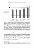
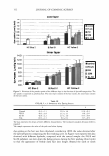
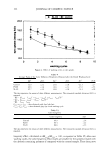
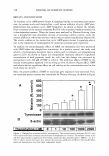
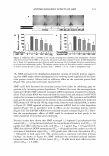
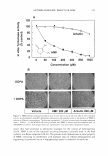
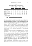
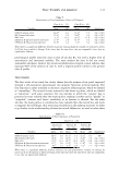
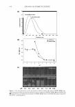
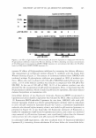
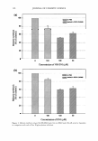
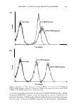
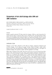
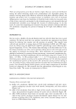
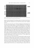
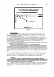
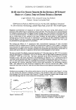
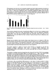
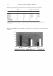
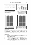
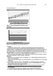
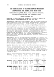
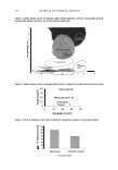
DELIVERY ACTIVITY OF pH-SENSITIVE LIPOSOMES 143 removal of the methanol, the slides were air-dried for 30 minutes and treated with Prolong Antifade solution (Molecular Probes, Eugene, OR) to prevent photobleaching of the fluorescent materials. The mounted slides were kept in the dark at 4°C until microscopic observation was performed. Cells were examined on a CLSM (Radiance 2000/MP, Bio-Rad, UK). RESULTS AND DISCUSSION N-glycosylation inhibitors were encapsulated in pH-sensitive liposomes consisting of DOPE and CHEMS to increase the depigmentation effect on HM3KO melanoma cells. It was evaluated that DOPE:CHEMS liposomes efficiently delivered whitening active molecules into the cytosolic active site adjacent to the endoplasmic reticulum (ER) of human melanoma cells. The size distribution of the N-glycosylation inhibitor-loaded pH-sensitive liposomes was examined by DLS, as shown in Figure la. The mean diam- eters of the prepared liposomes listed in Table I were in the range of 150-300 nm. The pH-sensitive characteristics of the DOPE:CHEMS liposomes were evaluated at various pH levels by turbidity measurements. The turbidity of the suspension containing DOPE:Chol (L6) liposomes was slightly increased between pH 6.0 and pH 5.0, and subsequently reached a plateau below pH 5.0 (Figure lb). While DOPE:Chol liposomes did not show conformational changes by decreasing pH, the turbidity ofDOPE:CHEMS (L1 ) was suddenly increased below pH 6.0, which is the early endosomal pH in cyto- plasm. Subsequently, the lipid components of pH-sensitive DOPE:CHEMS liposomes did not sustain a stable liposomal configuration. Thus, pH-sensitive liposomes collapsed below pH 5.0, a lower range of endosomal pH, and precipitated as shown in the lower part of Figure le. These results represents the pH sensitivity of DOPE:CHEMS lipo- somes, correlating with previous research (3,12). Note that CHEMS renders the vesicles pH-sensitive it stabilizes the lipid vesicles above neutral pH, while inducing structural instability via its own protonation at acidic pH. The cytotoxicity of pH-sensitive and pH-insensitive liposomes was evaluated using the MTT assay. Lipid concentrations of pH-sensitive liposomes and pH-insensitive liposomes up to 100 µM showed negligible cytotoxicity (data not shown). Therefore, 90 µM, the concentration of the lipid com- position, was used for the preparation of liposomes in all in vitro experiments. After incubation with HM3KO melanoma cells, the enhanced N-glycosylation- inhibiting (GI) effects of NB-DNJ or DNJ by the pH-sensitive delivery carrier were evaluated by Western blotting. Pigment-lightening effects were also monitored via the measurement of melanin biosynthesis by absorbence at 490 nm. When appropriate amounts of N-glycosylation inhibitors are delivered into melanocytes, the process of making a glycosylated tyrosinase (80 kDa) is inhibited and, thus, immature tyrosinase cannot help to synthesize the melanin (13). Therefore, the brighter band thickness of 80 kDa in the result of Western blotting meant that maturation of tyrosinase was inhibited and that the amount of a glycosylated tyrosinase (80 kDa) was decreased. Figure 2a shows that the GI effect of 50 µM of NB-DNJ-loaded pH-sensitive DOPE:CHEMS (L 1 ) liposome was similar to that of the 200 µM of intact NB-DNJ, by comparison of the band thickness of 80 kDa. Concentrations at 100 µM and even 50 µM of NB-DNJ and DNJ showed the enhanced GI effect by the delivery carrier, DOPE:CHEMS (L 1 ) lipo- somes, when it was compared to that of each 100 µM of free NB-DNJ and DNJ (Figure 26). Consequently, it could be demonstrated that the DOPE:CHEMS liposome can
Purchased for the exclusive use of nofirst nolast (unknown)
From: SCC Media Library & Resource Center (library.scconline.org)