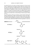
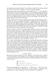
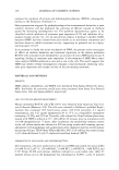
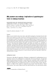
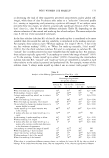
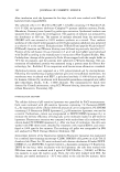
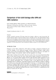
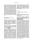
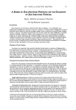
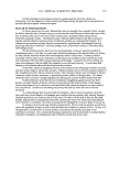
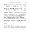
152 JOURNAL OF COSMETIC SCIENCE There are several amino acids that are brittle to light these are cystine and methionine among the sulfur-containing amino acids, phenylalanine and tryptophan among the aromatic and ring amino acids (which are associated with photo-yellowing of hairs), and histidine and proline (5 ,6). A gradual increase in brittleness and a loss of structural differentiation come from the breakdown of disulfide bonds within the structural units of the A-layer, exocuticle, and matrix. In the restoration process, new intramolecular and intermolecular crosslinks occur through the reaction of carbonyl groups (1). In this study, we carried out morphological evaluation and protein analysis of the hair shaft after UV irradiation, comparing the pattern of damage by UV light with different wave- lengths. EXPERIMENT AL For this study, a healthy 28-year-old Korean male was selected whose hair was in good condition. He had not used hair care products excessively nor experienced any factors known for inducing hair injuries over the previous six months. Hair samples were collected and irradiated to various doses of UVA (maximum 6,680]) and UVB (maxi- mum 58,320mJ) using the HOUVA II UAB-001 phototherapy system (National Bio- logical Corporation, U.S.A.). The distance from the lamps to the hair shafts was 50 cm, the relative humidity was 30%, and an external cooling device with cold water and ice was applied to minimize the heating effect. After irradiation, we accomplished mor- phological evaluation by scanning electron microscopy, conventional transmission elec- tron microscopy, and lipid transmission electron microscopy using Lee's fixative (0.5% RuO4 : 2% OsO4 : 0.2 M cacodylate buffer == 1 : 1 : 1) (7). Concurrently, we performed labile hair protein analysis according to the method of Inoue et al. (8), who have reported the transformation patterns of hair protein after permanent waving and hair dyeing in vitro. According to them, stable protein portions in normal hair are transformed to labile protein, the internally formed soluble protein, and the major components of the labile protein are ubiquitins (8). We proposed that this phenomenon would happen in irra- diated hair shafts. The results are summarized as follows. RESULTS AND DISCUSSION MORPHOLOGICAL FINDINGS USING ELECTRON MICROSCOPY Scanning electron microscopic study (a) Before UV irradiation: Normal hair cuticles look undamaged and have intact, tightly overlapping cuticular scales with smooth, homogenous, and shiny surfaces (Figure lA). (b) After UVA irradiation: Focal lifts at edges of the cuticles, focal losses of cuticular edges, and generalized irregular contours of cuticle cells are observed (Figure lB). (c) After UVB irradiation: Focal losses of cuticular edges, focal cuticular lifts at the edges of the cuticles, and focal cuticular detachments appear (Figure lC). In com- parison to findings after UV A irradiation, more severe cuticular damage seems to occur in DVB-irradiated hair shafts.

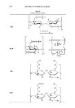
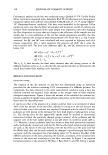
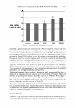
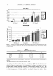
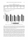
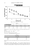
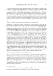
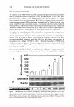
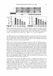
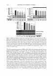
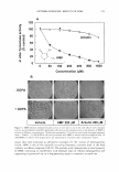
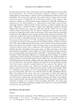
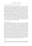
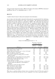
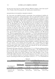
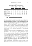
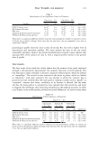
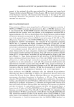
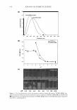
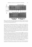
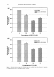
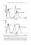
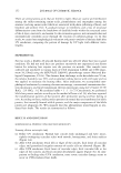
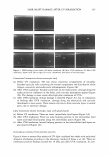
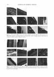
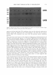
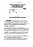
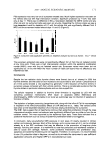
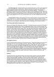
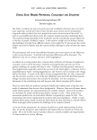
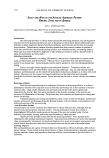
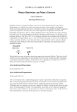
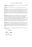
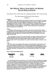
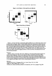
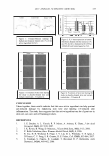
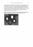
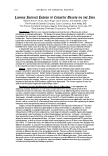
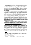
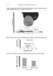
HAIR SHAFT DAMAGE AFTER UV IRRADIATION 153 Figure 1. SEM findings in hair shafts: (A) Before irradiation. (B) After UVA irradiation. (C) After UVB irradiation. Arrow: focal lifts at edges of cuticles. Arrowhead: focal losses of curicular edges. Conventional transmission electron microscopic study (a) Before UV irradiation: The hair shows concentric arrangements of smoothly bounded cuticular cells containing the normal complement and distribution of the A-layer, exocuticle, and endocuticle subcomponents (Figure 2A). (b) After UV A irradiation: Variable-sized holes in the endocuticles, cleavage along the endocuticles by confluence of the holes, and cuticular detachment appear (Figure 2B). The damage is more severe after high-dose irradiation of UVA. (c) After UVB irradiation: Similar damage is observed (Figure 2C), but in comparison to findings after UV A irradiation, cleavage along the endocuticle and cuticular detachment is more severe. There remain only two to three cuticular layers in several parts, due to extensive damage. Lipid transmission electron microscopic study with special fixative (a) Before UV irradiation: There are intact intercellular lipid layers (Figure 3A). (b) After UVA irradiation: There are some bulging portions in the intercellular lipid layers and small focal lacunae along the intercellular spaces (Figure 3B). (c) After UVB irradiation: Several bulging portions in the intercellular lipid layers are also observed (Figure 3C). BIOCHEMICAL FINDINGS WITH PROTEIN ANALYSIS Figure 4 shows a western blot analysis of UV-light-irradiated hair shafts with polyclonal ubiquitin antibodies according to the laboratory method of Inoue et al. (8). There are continuous positive findings around the 10 kDa area after UV A irradiation. In com-
Purchased for the exclusive use of nofirst nolast (unknown)
From: SCC Media Library & Resource Center (library.scconline.org)