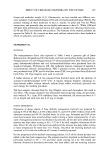
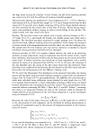
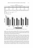
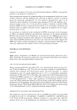
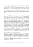
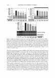
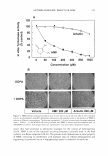
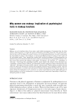
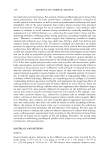
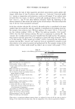
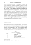
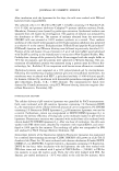
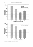
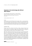
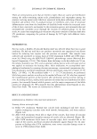
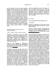
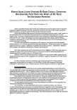
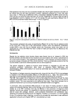
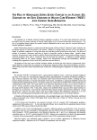
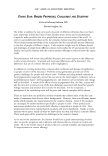
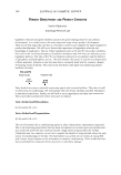
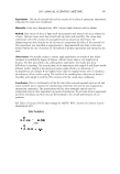
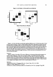
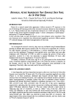
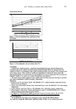
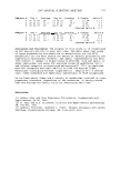
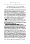
120 JOURNAL OF COSMETIC SCIENCE RESULTS AND DISCUSSION To stimulate cyclic AMP/protein kinase A signaling leading to tyrosinase gene expres- sion, the present study used theophylline, a well known inhibitor of cyclic AMP phos- phodiesterase that catalyzes cyclic AMP breakdown. As shown in Figure lA, theoph- ylline treatment of the murine melanoma B16F10 cells stimulated melanin synthesis in a dose-dependent manner. When the lysates were analyzed by Western blotting, there was a theophylline dose-dependent increase of tyrosinase protein content, while the content of �-actin, which was used as a control, was not altered significantly (Figure 1B). The results conform to the notion that cyclic AMP/protein kinase A signaling plays a role in the regulation of melanogenesis by inducing tyrosinase gene expression. To examine the antimelanogenic effects of HMF, the melanoma cells were pretreated with HMF before the theophylline stimulation. As a positive control, the study used arbutin, a hydroquinone derivative that is widely used in cosmetics as a depigmenting agent (12,18). As shown in Figure 2A, theophylline treatment induced a significant accumulation of dark pigments inside the cells, but this change was prevented by pretreatment with 200 µM of HMF or arbutin. The inhibitory effects of HMF on the cellular melanogenesis appeared to be as strong as those of arbutin (Figure 2B,C). HMF and arbutin had no significant effects on cell viability at the concentrations used in this study (data not shown). Next, the potential effects of HMF on tyrosinase gene expression were examined. First, the tyrosinase protein content was determined by Western blotting. As shown in Figure .-. 600 - A 0 500 .... C 0 400 u � 300 Cl ._. C 200 C ca 100 - CD ::il 0 Tyrosinase B B-actin I Theophylline (µM) I 0 * * * 1 1 1 o 11 oo 11 ooo 1 2000 1 Figure 1. Theophylline stimulates cellular melanogenesis. The cultured murine melanoma B16F10 cells were treated with vehicle or various concentrations of theophylline for 72 hours. The melanin content in the cell-free culture media is expressed as % of vehicle control (A). Data represent mean ± SEM (n = 3). *p 0.05 vs vehicle control. Cell lysates were analyzed for tyrosinase and (3-actin by Western blotting (B). Blots shown are representative of at least three independent studies.

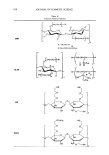
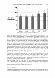
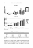
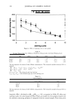
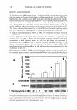
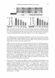
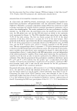
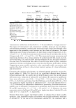
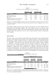
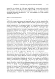
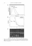
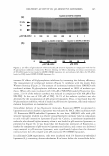
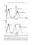
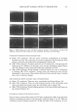
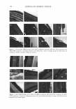
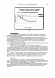
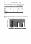
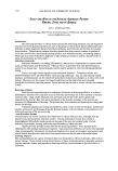
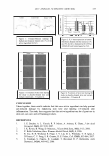
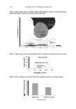
ANTIMELANOGENIC EFFECTS OF HMF 121 A Theophylline Figure 2. Inhibitory effects of HMF on the cellular melanogenesis stimulated by theophylline. B16F10 cells were pretreated with HMF or arbutin for 60 minutes and then stimulated with 1.0 mM theophylline for 72 hours. Cell pigmentation was observed under microscopy (A). Cell images shown are representative of at least three independent studies. The melanin content in the cell-free culture media is expressed as % of vehicle control (B and C). Data represent mean ± SEM (n = 3). *p 0.05 vs theophylline only. 3A, HMF attenuated the theophylline-dependent increase of tyrosine protein, suggest- ing that HMF might inhibit melanogenesis by involving down-regulation of the tyros- inase protein content. Arbutin had no inhibitory effect on the tyrosinase protein level (Figure 3B), as has been previously reported (18). HMF could have decreased the tyrosinase protein level either by inhibiting gene ex- pression or by increasing protein degradation. To address this issue, the next experiment examined whether HMF inhibited tyrosinase mRNA expression stimulated by theoph- ylline. Total cellular RNA was extracted from the treated cells and subjected to RT-PCR analysis for tyrosinase and housekeeping GAPDH mRNAs. The tyrosinase and GAPDH gene-specific primers designed in the present study successfully amplified the predicted PCR products of 564 bp and 396 bp, respectively, from the total cellular RNA, as shown in Figure 3C. HMF appeared to lower the tyrosinase mRNA level in a dose-dependent manner (Figure 3C) in agreement with its effects on the tyrosinase protein content (Figure 3A). The GAPDH mRNA level remained virtually constant (Figure 3C). There- fore the antimelanogenic effects of HMF could be attributed at least partly to the down-regulation of tyrosinase gene expression. Previous studies have shown that HMF scavenged 1,1-diphenyl-2-picrylhydrazyl free radicals (SC 50 = 3.0 µg/ml) and inhibited the autoxidation of linolenic acid (IC 9 0 = 32 µg/ml) more effectively than ascorbic acid (SC 50 = 3.3 µg/ml IC 90 = 124 µg/ml) (14). HMF also inhibited DOPA autoxidation (IC 50 = 60 µg/ml) and the catalytic activity of tyrosinase of mushroom origin (IC 50 = 100 µg/ml) more effectively than arbutin (IC50 300 µg/ml in both cases) (15). The current study is consistent with these previous findings. As shown in Figure 4A, HMF inhibited the tyrosinase-catalyzed melanin formation from DOPA in cell-free lysates (IC 50 = 50 µM) much more effectively than arbutin (IC50 1.6 mM).
Purchased for the exclusive use of nofirst nolast (unknown)
From: SCC Media Library & Resource Center (library.scconline.org)