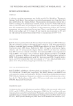
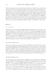
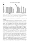
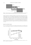
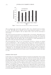
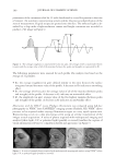
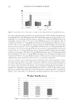
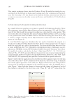
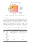
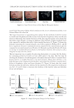
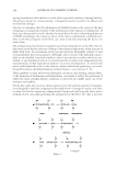
JOURNAL OF COSMETIC SCIENCE 154 since the skin comprises essentially three types of cells: keratinocytes, melanocytes, and fi broblasts. Given the importance of hyaluronan in the homeostasis of skin moisture, we next exam- ined the effect of PCS on hyaluronan synthesis in HaCaT cells. As a result, PCS remark- ably increased hyaluronan synthesis in HaCaT cells. Skin aging is closely associated with a loss of skin moisture. The key molecule involved in skin moisture is hyaluronan or hy- aluronic acid, a glycosaminoglycan (GAG) with a unique capacity to bind to and retain water molecules. Hyaluronan is present in many tissues and, in particular, is quite abun- dant in the matrix between keratinocytes, occurring predominantly in the spinous layer but also in the basal layer and stratum corneum. Hyaluronan is essential for maintaining moisture in skin tissues that contain approximately half of the body’s mass of hyaluronan. Third, elastase and procollagenase activities were examined in HDF-N cells. Moreover, since UVA radiation has been accepted as the most important cause of photoaging, charac- terized by alterations in the quantity of ECM, MMP activity was evaluated in UVA-exposed HDF-N cells. Our data showed that UVA-induced MMP activity was abrogated by PCS in a concentration-dependent manner. Procollagenase and elastase activities were decreased in HDF-N cells. Several lines of evidence have indicated a strong connection between the ac- tivity of dermal enzymes (collagenase and elastase) and wrinkle formation (31,32). Skin wrinkling, the appearance of visible signs on the skin surface (32), is a complex and inevi- table process of skin aging associated with an age-dependent decline in skin cell function. UVA radiation exposure to the skin results in the production of ROS implicated in MMP-1 activity responsible for collagen damage and photoaging. Indirect evidence sug- gests that 1 O2 and H2O2 are major ROS related to UVA-mediated activation of MMP-1, MMP-2, and MMP-3 (13,18,33). MMP-mediated ECM damage has been shown to be a major contributor of photoaging in human skin. For this reason, an antiwrinkle agent should have the potential to inhibit collagenase, elastase, and MMP activities (2,34). Next, to determine the whitening effects of PCS, a tyrosinase inhibition assay and melanin formation test were performed in Melan-a cells. Our results showed that PCS slightly Figure 4. Whitening effects of PCS. (A) Tyrosinase activity was assayed after KA (5 ppm) and PCS treat- ments (0.0125%, 0.025%, 0.05%, and 0.1%). (B) Melanin production was determined following treatment with arbutin (100 μM) and PCS (0.005%, 0.01%, and 0.05%) for 72 h. Values are expressed as the means ± S.D. from fi ve independent experiments. a p 0.01 compared with the control (MW test).

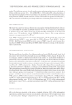
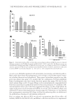
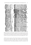
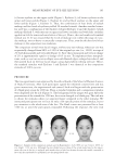
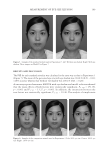
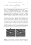
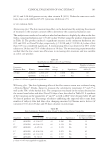
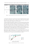
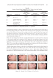
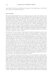
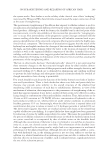
THE WHITENING AND ANTI-WRINKLE EFFECT OF PONEGRANATE 155 inhibited tyrosinase activation, whereas melanin formation was signifi cantly inhibited by 0.01% and 0.05% PCS. These results suggest that PCS may affect skin whitening. Immortalized mouse melanocytes, Melan-a cells, were derived from normal epidermal melanoblasts from embryos of inbred C57BL mice. Thus, Melan-a cell line was used to evaluate the effects of skin whitening. Melanin is produced in melanocytes and Figure 5. Representative histological images of dorsal back skin tissues taken from unexposed intact or UVB-exposed hairless mice. (A) Normal, nonirradiated vehicle control hairless mice (intact control). (B) UVB-irradiated vehicle control hairless mice (UVB control). (C) UVB-irradiated and PCS-administrated hairless mice (PCS-0.5). (D) UVB-irradiated and PCS-treated hairless mice (PCS-1). (E) UVB-irradiated and PCS-administrated hairless mice (PCS-2). (F) Normal, nonirradiated and PCS-administrated hairless mice (intact PCS-1). Arrows indicate microfolds formed. All tissues were stained with H&E. Scale bar = 50 μm.
Purchased for the exclusive use of nofirst nolast (unknown)
From: SCC Media Library & Resource Center (library.scconline.org)