159 Hydrolyzed Conchiolin Protein Inhibits Melanin
have been employed for skin whitening by inhibiting melanin production, there is still a
lack of drugs that specifically target melanin transfer. Current whitening compounds, such
as hydroquinone and alpha arbutin, have been reported to be carcinogenic and can cause
redness or irritation when used in large amounts.9 Therefore, there has been an increasing
emphasis on the development of safer whitening products.
Pearl extract is rich in minerals and amino acids. Pearl extracts can enhance skin moisture, skin
repair, and UV protection without any unwanted side effects. It has been demonstrated that a
soluble pearl extract possesses the ability to inhibit melanin synthesis in B16 melanoma cells.10
In vitro studies have also shown that HCP exhibits anti–inflammatory and anti–apoptotic
properties.11 HCP is a hydrolyzing derivative of conchiolin protein extracted from pearls. As
a significant active component of pearl extract, HCP has been found to be biocompatible, but
the specific effects of HCP on melanin synthesis and transport remain unknown.
In this study, we aimed to investigate the effects of HCP on melanocyte morphology and
melanosome transport, and to further elucidate the underlying mechanism. We observed
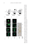
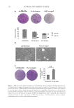
that HCP effectively suppressed melanocyte dendrite extension induced by ET-1 and
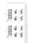
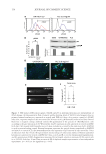
downregulated the expression of genes involved in melanosome transport, including MITF,
Rab27A, and Rac1, suggesting a novel role for HCP in regulating melanosome transport.
Additionally, HCP was found to negatively regulate the activity of the Wnt/β-catenin
signaling pathway, leading to the decrease of pigmentation in zebrafish. Therefore, it seems
that HCP can inhibit melanin transfer by suppressing the activity of Wnt/β-Catenin
signaling pathway in melanocytes.
MATERIALS AND METHODS
REAGENTS
HCP was obtained from OSM Biology Co., Ltd. (Zhejiang, China). ET-1 was purchased
from MedChemExpress Co., Ltd. (Shanghai, China).
CELL CULTURE
PIG1 and MelanA cells were purchased from American Type Culture Collection (ATTC,
Virginia, USA) and cultured at 37°C with 5% CO
2 atmosphere in Dulbecco’s Modified
Eagle Medium (DMEM) containing 10% fetal bovine serum, 100 U/ml penicillin, and
100 µg/ml streptomycin.
CELL VIABILITY ASSAY
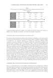
PIG1 and MelanA cells were cultured in 96-well plates at a density of 1.5 × 10⁵/ml. Cells
were treated with HCP and ET-1 at various concentrations for 24 hours. For PIG1 cells, we
selected HCP treatment concentrations of 0, 0.01, 0.1, 5, 10, 20, 50, 80, and 100 µg/ml.
For MelanA cells, we selected HCP treatment concentrations of 0, 0.1, 1, 2, 4, 8, 10,
20, 40, 80 µg/ml. After a brief wash with medium, 3-(4,5-dimethylthiazol-2-yl)-5-(3-
carboxymethoxyphenyl)-2-(4-sulfophenyl)-2H-tetrazolium (MTS) (0.5 mg/ml in DMEM)
was used for the quantification of metabolically active cells, and samples were measured
photometrically at 490 nm.
have been employed for skin whitening by inhibiting melanin production, there is still a
lack of drugs that specifically target melanin transfer. Current whitening compounds, such
as hydroquinone and alpha arbutin, have been reported to be carcinogenic and can cause
redness or irritation when used in large amounts.9 Therefore, there has been an increasing
emphasis on the development of safer whitening products.
Pearl extract is rich in minerals and amino acids. Pearl extracts can enhance skin moisture, skin
repair, and UV protection without any unwanted side effects. It has been demonstrated that a
soluble pearl extract possesses the ability to inhibit melanin synthesis in B16 melanoma cells.10
In vitro studies have also shown that HCP exhibits anti–inflammatory and anti–apoptotic
properties.11 HCP is a hydrolyzing derivative of conchiolin protein extracted from pearls. As
a significant active component of pearl extract, HCP has been found to be biocompatible, but
the specific effects of HCP on melanin synthesis and transport remain unknown.
In this study, we aimed to investigate the effects of HCP on melanocyte morphology and
melanosome transport, and to further elucidate the underlying mechanism. We observed
that HCP effectively suppressed melanocyte dendrite extension induced by ET-1 and
downregulated the expression of genes involved in melanosome transport, including MITF,
Rab27A, and Rac1, suggesting a novel role for HCP in regulating melanosome transport.
Additionally, HCP was found to negatively regulate the activity of the Wnt/β-catenin
signaling pathway, leading to the decrease of pigmentation in zebrafish. Therefore, it seems
that HCP can inhibit melanin transfer by suppressing the activity of Wnt/β-Catenin
signaling pathway in melanocytes.
MATERIALS AND METHODS
REAGENTS
HCP was obtained from OSM Biology Co., Ltd. (Zhejiang, China). ET-1 was purchased
from MedChemExpress Co., Ltd. (Shanghai, China).
CELL CULTURE
PIG1 and MelanA cells were purchased from American Type Culture Collection (ATTC,
Virginia, USA) and cultured at 37°C with 5% CO
2 atmosphere in Dulbecco’s Modified
Eagle Medium (DMEM) containing 10% fetal bovine serum, 100 U/ml penicillin, and
100 µg/ml streptomycin.
CELL VIABILITY ASSAY
PIG1 and MelanA cells were cultured in 96-well plates at a density of 1.5 × 10⁵/ml. Cells
were treated with HCP and ET-1 at various concentrations for 24 hours. For PIG1 cells, we
selected HCP treatment concentrations of 0, 0.01, 0.1, 5, 10, 20, 50, 80, and 100 µg/ml.
For MelanA cells, we selected HCP treatment concentrations of 0, 0.1, 1, 2, 4, 8, 10,
20, 40, 80 µg/ml. After a brief wash with medium, 3-(4,5-dimethylthiazol-2-yl)-5-(3-
carboxymethoxyphenyl)-2-(4-sulfophenyl)-2H-tetrazolium (MTS) (0.5 mg/ml in DMEM)
was used for the quantification of metabolically active cells, and samples were measured
photometrically at 490 nm.