174 JOURNAL OF COSMETIC SCIENCE
increasing the risk of genetic alterations. Hence, skin protection against UV light can be
considered preventive medicine, as there becomes a need toward preventing short-term
(erythema, inflammation) and long-term (cancer, aging) effects of exposure to this harmful
radiation. Epidermal keratinocytes are a major target of radiation-induced damage as they
make up most epidermal cells.
In the last decades, the public has become widely aware of the risks of sustained exposure
to sunlight, and the use of sun-protection products has significantly increased. Diverse
natural products have been used to counteract UV radiation–induced skin damage
through different approaches. One is the use of sunscreens, which prevent high-energy
photons from reaching sensitive biological targets, e.g., nuclear DNA these substances
possess the ability to absorb photonic energy, thereby preventing biological substrates
from doing it. Another possibility is to use substances that do not act as a sunscreen
but minimize injury though alternative mechanisms, e.g., attenuating oxidative or DNA
damage. These substances do not necessarily need to be applied topically, but they can
also be administered orally.5
Our group has focused on the beneficial effects of a hydrophilic extract from the leaves
of the fern Polypodium leucotomos (PLE, Fernblock®). PLE has a remarkable safety profile
(Nestor et al., 2015), has a function as a barrier (it absorbs UV photons), and acts as
a biological modifier.6,7 However, its main beneficial effect is due to its extraordinary
antioxidant ability and protective cellular effects. These include improved myeloid
(Langerhans) cell survival upon UV irradiation, decreased trans–urocanic acid
isomerization and increased DNA repair enzyme activity.8–11 PLE is currently used in
topical and oral sunscreen formulations.12 It also counteracts other deleterious effects of
UV irradiation, e.g., photoaging and photoimmunosuppression.13,14 Its mechanism of
action seems multipronged, but its antioxidant capability plays a fundamental role in its
beneficial effects.9
Although the above beneficial effects are well documented, the genetic protective
mechanisms of PLE in the keratinocyte biology have not been addressed yet. Unrepaired
DNA damage blocks proliferation and induces differentiation of keratinocytes of the diverse
stratified epithelia of the skin or head and neck.15–17 This response is controlled by mitotic
checkpoints.16,18–20 The differentiation response protects keratinocytes from apoptosis even
upon moderate UV irradiation.21 The G2 phase of the cell cycle, prior to mitosis, is a major
phase for DNA repair. Upon DNA damage, G2 checkpoints halt the cycle to ensure that
DNA is repaired before cell division.22 Multipotent stem cells within the basal layer of the
epidermis maintain the tissue and are targets of genetic insult and aging.23,24 These cells
are not totipotent but are capable of replenishing the tissue and giving rise to different
progenitors.
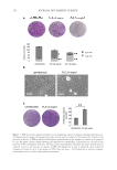
We have investigated the effects of treating primary human keratinocytes with PLE on
DNA damage and the cell cycle before or after UV irradiation. Interestingly, epidermal
keratinocytes treated with PLE were retarded in growth but conserved a greater capacity to
amplify after treatment. We have investigated the molecular mechanisms underlying this
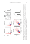
protective effect. A “soft arrest” in G2/M upon PLE treatment might allow cells to better
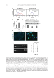
repair their DNA before undergoing mitosis. PLE appears to achieve this by enhancing
cellular DNA repair signals. This mechanism might contribute to preserve genome
integrity and the proprieties of epidermal stem cells face to DNA replication stress or UV
irradiation. We discuss the implications for skin cancer and aging.
increasing the risk of genetic alterations. Hence, skin protection against UV light can be
considered preventive medicine, as there becomes a need toward preventing short-term
(erythema, inflammation) and long-term (cancer, aging) effects of exposure to this harmful
radiation. Epidermal keratinocytes are a major target of radiation-induced damage as they
make up most epidermal cells.
In the last decades, the public has become widely aware of the risks of sustained exposure
to sunlight, and the use of sun-protection products has significantly increased. Diverse
natural products have been used to counteract UV radiation–induced skin damage
through different approaches. One is the use of sunscreens, which prevent high-energy
photons from reaching sensitive biological targets, e.g., nuclear DNA these substances
possess the ability to absorb photonic energy, thereby preventing biological substrates
from doing it. Another possibility is to use substances that do not act as a sunscreen
but minimize injury though alternative mechanisms, e.g., attenuating oxidative or DNA
damage. These substances do not necessarily need to be applied topically, but they can
also be administered orally.5
Our group has focused on the beneficial effects of a hydrophilic extract from the leaves
of the fern Polypodium leucotomos (PLE, Fernblock®). PLE has a remarkable safety profile
(Nestor et al., 2015), has a function as a barrier (it absorbs UV photons), and acts as
a biological modifier.6,7 However, its main beneficial effect is due to its extraordinary
antioxidant ability and protective cellular effects. These include improved myeloid
(Langerhans) cell survival upon UV irradiation, decreased trans–urocanic acid
isomerization and increased DNA repair enzyme activity.8–11 PLE is currently used in
topical and oral sunscreen formulations.12 It also counteracts other deleterious effects of
UV irradiation, e.g., photoaging and photoimmunosuppression.13,14 Its mechanism of
action seems multipronged, but its antioxidant capability plays a fundamental role in its
beneficial effects.9
Although the above beneficial effects are well documented, the genetic protective
mechanisms of PLE in the keratinocyte biology have not been addressed yet. Unrepaired
DNA damage blocks proliferation and induces differentiation of keratinocytes of the diverse
stratified epithelia of the skin or head and neck.15–17 This response is controlled by mitotic
checkpoints.16,18–20 The differentiation response protects keratinocytes from apoptosis even
upon moderate UV irradiation.21 The G2 phase of the cell cycle, prior to mitosis, is a major
phase for DNA repair. Upon DNA damage, G2 checkpoints halt the cycle to ensure that
DNA is repaired before cell division.22 Multipotent stem cells within the basal layer of the
epidermis maintain the tissue and are targets of genetic insult and aging.23,24 These cells
are not totipotent but are capable of replenishing the tissue and giving rise to different
progenitors.
We have investigated the effects of treating primary human keratinocytes with PLE on
DNA damage and the cell cycle before or after UV irradiation. Interestingly, epidermal
keratinocytes treated with PLE were retarded in growth but conserved a greater capacity to
amplify after treatment. We have investigated the molecular mechanisms underlying this
protective effect. A “soft arrest” in G2/M upon PLE treatment might allow cells to better
repair their DNA before undergoing mitosis. PLE appears to achieve this by enhancing
cellular DNA repair signals. This mechanism might contribute to preserve genome
integrity and the proprieties of epidermal stem cells face to DNA replication stress or UV
irradiation. We discuss the implications for skin cancer and aging.