182 JOURNAL OF COSMETIC SCIENCE
PLE ATTENUATES THE DIFFERENTIATION RESPONSE TO DNA DAMAGE
Epidermal stem cells are able to give rise to various linages and have a great capacity of
amplification in vitro.1,23,25,35 Epidermal stem cells are targets of genetic damage and aging.
Allowing more time in the DNA repair G2 phase should prolong the renewal capacity of
stem cells, and this is what we observed when cells were released after PLE treatment. The
boost of the DNA repair signal also should diminish the proportion of cells undergoing
differentiation postmitotically. Premature keratinocyte differentiation eventually
contributes to skin aging. Cells treated with PLE were more protected against aging, as they
retained a greater capacity for self-renewal and amplification after treatment. Consistently,
we observed an attenuated rate of terminal differentiation in both exponentially growing
cells and UV-irradiated cells that were treated with PLE. UV light and aging are the main
causes of skin epithelial cancer. The mechanism we describe might support the beneficial
anticancer activity of the PLE in response to UV radiation.5
A MECHANISM POTENTIALLY COMMON TO OTHER SOLAR PROTECTIVE COMPOUNDS
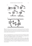
The model we draw might be common to other natural substances protecting cells from
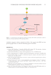
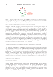
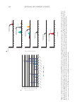
UV light (Figure 6). This is in line with observations that many antioxidant agents inhibit
cell proliferation in a concentration-dependent manner, e.g., N-acetyl cysteine or green tea
polyphenols.36,37 The effects on the cell cycle described here show that PLE has a multipronged
function in protecting cellular DNA by controlling the cell cycle and favoring repair and
checkpoint assessment. Future studies will elucidate the specific mechanisms and signaling
cascades by which PLE and possibly other natural substances protect the epidermal cell cycle.
MATERIALS AND METHODS
KERATINOCYTE CULTURE
Primary human keratinocytes were isolated from neonatal foreskins of two different
individuals and cultured Rheinwald conditions in the presence of a fibroblastic feeder
layer of J2-3T3 cells (inactivated by 6 µg/ml mitomycin C) in 5% fetal calf serum–FAD
medium (1,6 mM — “high”— calcium), as described previously 4 × 10⁵ cells were plated
per 10-cm dish. J2-3T3 cells were cultured in DMEM containing 10% calf serum, 2 mM
L-Glutamine, and 100 U/mL penicillin/streptomycin.25,35
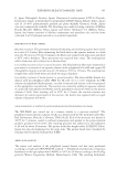
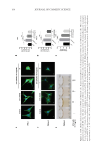
Figure 6. Proposed model for the protective effect of PLE on the epidermal cell cycle and renewal. By
slowing down the cell cycle at the G2 phase, PLE provides keratinocytes with more time to achieve DNA
repair, therefore protecting the integrity and renewal capacity of stem cells.
PLE ATTENUATES THE DIFFERENTIATION RESPONSE TO DNA DAMAGE
Epidermal stem cells are able to give rise to various linages and have a great capacity of
amplification in vitro.1,23,25,35 Epidermal stem cells are targets of genetic damage and aging.
Allowing more time in the DNA repair G2 phase should prolong the renewal capacity of
stem cells, and this is what we observed when cells were released after PLE treatment. The
boost of the DNA repair signal also should diminish the proportion of cells undergoing
differentiation postmitotically. Premature keratinocyte differentiation eventually
contributes to skin aging. Cells treated with PLE were more protected against aging, as they
retained a greater capacity for self-renewal and amplification after treatment. Consistently,
we observed an attenuated rate of terminal differentiation in both exponentially growing
cells and UV-irradiated cells that were treated with PLE. UV light and aging are the main
causes of skin epithelial cancer. The mechanism we describe might support the beneficial
anticancer activity of the PLE in response to UV radiation.5
A MECHANISM POTENTIALLY COMMON TO OTHER SOLAR PROTECTIVE COMPOUNDS
The model we draw might be common to other natural substances protecting cells from
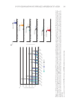
UV light (Figure 6). This is in line with observations that many antioxidant agents inhibit
cell proliferation in a concentration-dependent manner, e.g., N-acetyl cysteine or green tea
polyphenols.36,37 The effects on the cell cycle described here show that PLE has a multipronged
function in protecting cellular DNA by controlling the cell cycle and favoring repair and
checkpoint assessment. Future studies will elucidate the specific mechanisms and signaling
cascades by which PLE and possibly other natural substances protect the epidermal cell cycle.
MATERIALS AND METHODS
KERATINOCYTE CULTURE
Primary human keratinocytes were isolated from neonatal foreskins of two different
individuals and cultured Rheinwald conditions in the presence of a fibroblastic feeder
layer of J2-3T3 cells (inactivated by 6 µg/ml mitomycin C) in 5% fetal calf serum–FAD
medium (1,6 mM — “high”— calcium), as described previously 4 × 10⁵ cells were plated
per 10-cm dish. J2-3T3 cells were cultured in DMEM containing 10% calf serum, 2 mM
L-Glutamine, and 100 U/mL penicillin/streptomycin.25,35
Figure 6. Proposed model for the protective effect of PLE on the epidermal cell cycle and renewal. By
slowing down the cell cycle at the G2 phase, PLE provides keratinocytes with more time to achieve DNA
repair, therefore protecting the integrity and renewal capacity of stem cells.