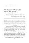
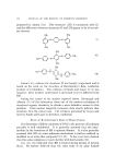
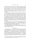
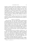
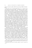
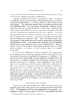
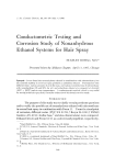
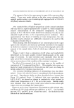
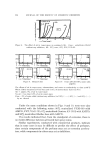
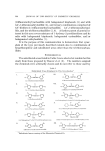
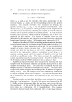
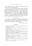
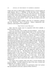
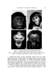
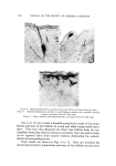
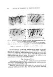
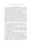
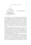
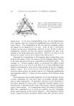
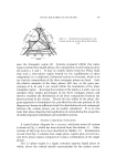
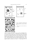
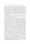
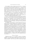
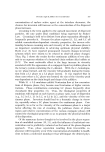
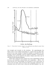
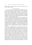
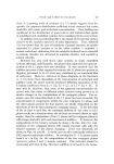
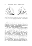
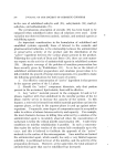
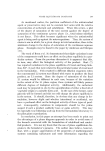
BALDNESS IN NONHUMAN PRIMATES 179 THE HAIR FOLLICLES Hair follicles generally grow in groups of two or three, with one apocrine sweat gland associated with each group on the face the follicles usually grow singly. In those scalp areas that are still covered with long hairs, the groups consist of one or two large terminal and one or two vellus hairs. The bald forehead and scalp are covered with a sparse population of vellus hairs with an occasional terminal hair scattered among them. Although the follicles in the bald areas are notably re- duced in size, most still have arrectores pilorum muscles attached to them true veilus follicles have no muscle. Terminal hair follicles have a small bulge for the attachment of stout muscles. In quiescent "veilus" follicles the muscle, when present, is attached to the epithelial capsule around the club hair, below which is the small hair germ. Quiescent terminal follicles are about one-half the length of the growing ones, the small, eccentric hair germ extending only a short distance below the bulge. Sinus hair follicles are found in the median aspect of the eyebrows, on the mystacial areas, on the lower lip, and on the chin scattered among the follicles of the goatee. Although grossly the transitional area between the bald and nonbald portions of the scalp appears to be abrupt, in histological sections it is surprisingly gradual. The normal-sized terminal hair follicles grow sparsely, having among them numerous veilus types or intermediate follicles. Farther back, on the temporal and occipital areas, where the terminal hairs grow longest, there is an appreciable population of small follicles. Even on the back of the trunk and nape, where the hairs form the mane, and elsewhere nearly every hair group contains small ones. In the scalp and face every follicle, large or small, has a complex of sensory nerves that compose the follicle nerve end-organ (Fig. 6). This is found around the pilary canal between the bulge and duct of the seba- ceous glands. These nerves, reactive for both butyryl- and acetyl- cholinesterase, are arranged in both a perpendicular and a horizontal direction. Elsewhere, not all of the follicles have such an abundant nerve supply. The sinus hair follicles are enveloped by very fine nerves that swirl around the upper portion, just below the sebaceous glands. In the forehead and eyebrows, the bald areas of the scalp and in some parts of the naked face, cholinesterase-containing nerve fibers are found in the papillary layer of the dermis that accompanies the underside of the epidermis for some distance. These terminate at the base of some of the epidermal ridges in small bulbous endings or in small bodies that re- semble mucocutaneous end-organs (Figs. 7, 8).

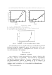
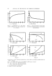
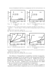
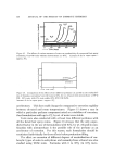
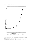
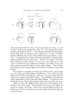
180 JOURNAL OF THE SOCIETY OF COSMETIC CHEMISTS Figure G. Butyrylcholinesterase activity in the hair follicle end-organ from the scalp Ftgure 7. Butyrylcholinesterase activity in small bulbous endings that resemble mucocu- tancous endorgans from the bald scalp Figure 8. More extensive mucocutaneous-like end-organs from the bald scalp Uno et al. (3) have made a detailed quantitative study of the popu- lations and sizes of hair follicles in young and adult stump-tailed maca- ques. They have also dissected out intact hair follicles from the sur- rounding connecting tissue to measure accurately their size and to study (to be reported later) their enzyme content, eliminating the contam- ination of surrounding tissues. Their results are shown in Figs. 9 to 12. Here are recorded the actual representative populations and sizes of hair follicles on the lower
Purchased for the exclusive use of nofirst nolast (unknown)
From: SCC Media Library & Resource Center (library.scconline.org)