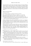
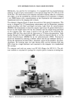
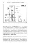
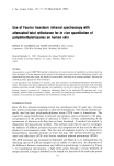
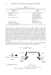
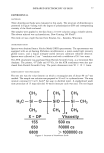
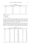
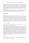
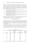
INFRARED SPECTROSCOPY OF SKIN 77 EXPERIMENTAL MATERIALS Three dimethicone fluids were evaluated in this study. The structure of dimethicone is presented in Figure 4 along with the degree of polymerization (DP) and corresponding viscosity of the fluids evaluated. The samples were applied to the skin from a 10 wt% solution using a volatile solvent. The solvent selected was cyclomethicone, Dow Corning 344 Fluid ©. The mink oil was a light fraction from Emulan, Inc., Kenosha, Wisconsin. INSTRUMENTATION Spectra were obtained from a Nicolet Model 20DX spectrometer. The spectrometer was equipped with an air-bearing Michelson interferometer, a water-cooled high intensity globar source, and a liquid nitrogen-cooled mercury cadmium telluride detector. Spectra were collected at 2 cm-1 resolution and with co-addition of 50 1-sec scans. The ATR attachment was purchased from Harrick Scientific Corp. as a horizontal Skin Analyzer. The prisms, 45ø-ZnSe and 45ø-Ge, for the ATR attachment were also pur- chased from Harrick Scientific Corp. The prism dimensions were 50 x 10 x 3mm. SUBSTANTIVITY TEST PROCEDURE The test site was the volar forearm on which a rectangular area of about 80 cm 2 was marked. The sample test solution was prepared at 10 wt% in cyclomethicone. The soap mixture contained 0.5 wt% Ivory © bar soap in distilled water. A standardized wash/ rinse procedure of 15 soap rubs and 10 water rinses per cycle was used. The test began CH 3 I H3C•Si•O I CH3 CH3 I sio I CH 3 - - X X - DP Viscosity 155 500 cs. CH 3 I Si•CH 3 I CH3 650 30000 cs 6800 1000000 cs Figure 4. Structure of dimethicone fluids.

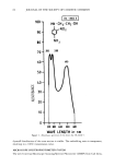
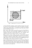
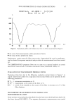
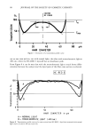
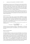
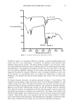
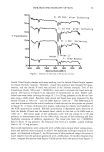
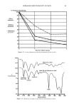
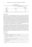
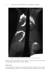
78 JOURNAL OF THE SOCIETY OF COSMETIC CHEMISTS by soap washing and rinsing the test site. We blotted dry the area, waited one minute, and softened the site for one minute with a water-soaked towel. We blotted dry the area and within 30 seconds placed the test site on the ATR prism and collected a back- ground scan of the skin surface. We applied a known, weighed quantity of test solution on the skin. Generally, a brush was used to apply the solution. The quantity applied was calculated by weight difference of solution vial plus brush before and after applica- tion of solution on the skin. A maximum loading of 10-12 mg of the non-volatile material on the 80 cm 2 area was a reasonable working limit. We waited 15-30 minutes for solvent evaporation. We softened the test site and col- lected an IR scan for the initial condition. We soap washed, rinsed, blotted dry, soft- ened, and collected IR scans for as many soap wash cycles as were of interest. Three soap wash cycles were generally used. We proceeded to the calculation procedures for quan- titation of material on skin. QUANTITATION PROCEDURE Within the Nicolet computer system, individual programs (MACROs) can be devel- oped to automatically calculate band absorbances or areas of specific bands. MACROs were developed to calculate the data for the SiMe band at 1260 cm- 1 and the Amide II protein band at 1540 cm-1 with respect to specific baselines drawn for each band. The baselines used with the Ge prism were 1780-1487 cm-1 for the Amide II band and 1352- 1198 cm- 1 for the SiMe band. This procedure simulates standard IR calculation procedures and eliminates hand calculations. Substantivity data was calculated as percentage remaining after each soap wash cycle using Equation [2]. % Remaining = [A3/A2] CONDITION X 100 [2] [A3/A 2] INITIAL In Equation [2], A 3 is the band area or absorbance of SiMe at 1260 cm-• and A2 is the band area or absorbance of Amide II at 1540 cm-•. The INITIAL value is calculated after application of test material. The CONDITION value is calculated after each soap wash cycle. RESULTS AND DISCUSSION METHOD DEVELOPMENT Prism Selection. Many prism materials can be used with ATR analysis. Two prism mate- rials commonly used for in vivo human skin studies are Ge and ZnSe. Both Ge and ZnSe are non-toxic, water resistant, and have acceptable IR detection ranges. The spectra presented in Figure 5 are characteristic of skin. A vertical displacement of some of the spectra presented in this paper was necessary for easy comparison of different conditions. Thus, the y-axis may indicate greater than 100% transmittance for some examples. The Amide I band at 1650 cm-• is predominantly due to carbonyl stretch. The Amide II band at 1540 cm-• is due predominantly to NH deformations. The increased overall intensity of the spectrum with the ZnSe prism, as compared to the Ge prism spectrum, is due to the greater penetration depth of the radiation caused by refractive index differ- ences between skin and the prism. Either prism can be used for skin studies.
Purchased for the exclusive use of nofirst nolast (unknown)
From: SCC Media Library & Resource Center (library.scconline.org)