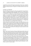
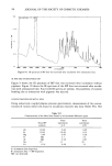
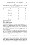
110 JOURNAL OF THE SOCIETY OF COSMETIC CHEMISTS rial. As others have reported (2,4), Quatrale et al. (7) also found that the site of action for ACH was primarily at the level of the stratum corneum compared to a deeper site for A1C13. The presence of aluminum at sites of pore blockage has been demonstrated (4-6), but additional chemical characterization of the inhibited lumen has been speculative at best. How and under what conditions precipitation is favored are factors that remain unclear. Characterizing the contents of an inhibited sweat duct is a first step in answering mech- anistic questions not only for aluminum salts but perhaps for all metal salt antiper- spirants. We have revealed and isolated these minute plug particulates from sweat glands of human forearm inhibited by ACH. With the high energy throughput of the FTIR spectrometer, a beam condenser, and a high pressure diamond anvil cell, we have been able to obtain infrared spectra of human biopsy plug material. We present new infor- mation on the nature of the area of sweat inhibition and hence what may be another significant factor in the mechanism of antiperspirant action. In addition, in vitro experiments were performed to find and characterize ACH-protein coordination sites and to aid in characterizing the in vivo plug spectra. Interactions were anticipated because AI sorption studies performed on guinea pig stratum corneum showed strong binding between these species as determined by bath depletion methods (8). EXPERIMENTAL MATERIALS Stratum corneum and sweat duct material (plugs) were obtained from full-thickness punch biopsies of human male forearm and axilla. For reference, infrared spectra were taken on four other human proteins which could conceivably be contained in a sweat duct. Three were obtained from U.S. Biochemicals (Cleveland, OH), while a fourth was isolated from the upper body (back and chest) perspiration of two thermally stressed males. The description of these proteins is given in Table I. Twenty percent aluminum chlorohydrate (ACH) solutions used for in vitro studies were diluted from 50% solutions (Reheis Chemicals, Phoenix, AZ, Batch #6085). This was a 5/6 basic ACH having an AI analysis of 12.35% w/w. Sodium chloride was A.C.S. certified reagent grade (Fisher Scientific Company, Fairlawn, N J). Fresh preparations of ACH were also made from a 50% solution (Westwood Chemical Company, Lot #PBC 03279). INSTRUMENTATION Infrared spectra were made on a Digilab FTS-10M Fourier transform infrared spectrom- Table I Proteins Used for FTIR Analysis 1. Glycoprotein, Fraction VI Human, Control No. 22777 2. ot-Globulins, Fraction IV Human, Control No. 12823 3. Albumin, Crystallized Human, Control No. 15153 4. Human Sweat--Centrifuged, filtered through 0.22 •xm Falcon sterile filter, dialyzed overnight at 3øC, and lyophilized to dryness

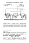
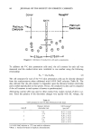
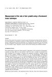
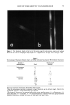
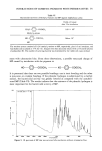
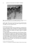
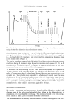
FTIR OF SWEAT GLANDS & ALUMINUM SALTS ! 11 eter purged with dry nitrogen and equipped with a hot wire source and a triglycine sulfate detector. Spectra were recorded at 4 cm-• resolution with the use of boxcar apodization, and 1000 scans were signal averaged for each spectrum to attain a signal- to-noise ratio yielding good spectral subtraction conditions. Two microsampling techniques were used for all studies with modifications when needed. A 6X beam condenser was used to concentrate the beam to about 2 mm diam- eter at the focal point. The first technique utilized a high pressure diamond anvil cell (High Pressure Diamond Optics, Tucson, AZ) in which the sample was pressed be- tween the two polished diamond faces. The diamonds, mounted on pistons, were squeezed by the lever action of a spring on the pistons. The pressure was cycled to spread the sample, and spectra were recorded at zero pressure with the spring loosened. A full description of the diamond cell is given elsewhere (9). Where larger samples were available, they were flattened between two gold mesh screen grids (of the type used for electron microscopy) and masked on the periphery with larger (1 x 2 mm) crossed grids. The grids and sample were mounted in a specially fabricated microsample holder, which likewise held the sample at the focal point of the beam condenser. Reference spectra were generated with either a clean diamond cell or empty grids in the 6X beam condenser. Sample holes in the infrared windows were masked to minimize spectral dilution or fringing effects. ISOLATION OF HUMAN ECCRINE SWEAT GLANDS Biopsies (!) 2 mm) were taken from the volar forearms of two human males. One forearm was treated overnight with an occlusive patch of 20% ACH the other arm had no treatment. The following day the subjects were thermally stressed at 100øF and 35% RH and biopsies taken at sites devoid of sweating. The biopsies were preserved under liquid nitrogen and brought to room temperature for dissection. They were fixed for two days at 4øC in 6.25% glutaraldehyde buffered with 0.1M Sorensen's phosphate buffer, pH 7.3. Each biopsy specimen was sliced with a razor blade into 0.2-mm sections parallel to several sweat glands. Immersion in glycerine revealed the gross anatomy of the sweat gland with a ductal area substantially darker (under transmitted light) than any surrounding tissue. The dark material was considered to be the region of poral occlusion or a plug from an ACH-inhibited sweat duct. Sections were also cut to demonstrate sweat ducts from the untreated forearm where no dark material was found. A sliced section is shown in Figure !. The dark plug material (usually found near the stratum corneum/viable epidermis interface) was gently removed from the surrounding tissue with specially ground, fine-tipped tweezers. It was not possible to remove cleanly the dark plug material without drawing a small amount of surrounding tissue with it. Approximately three or four excised plugs were used to compose each of the samples designated as plug # ! and #2 and taken from different subjects. Additional stratum corneum was dissected from the same ACH-treated biopsy as plug #1 but about 2 mm away from the sweat duct. A control sample was sectioned from the biopsy from the untreated forearm and dissected to contain part of the sweat duct as well as surrounding stratum corneum. All FTIR analyses in these experiments were carried out using the high pressure diamond anvil cell. Although the plug samples were thinned considerably in the cell, they were not large enough to completely fill the diamond windows (--1 mm2). The beam was partially masked with an iris to minimize spectral dilution effects. The spectral quality from plug #2 was superior to that from
Purchased for the exclusive use of nofirst nolast (unknown)
From: SCC Media Library & Resource Center (library.scconline.org)