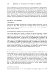
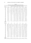
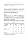
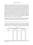
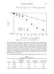
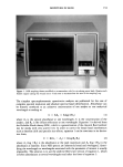
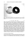
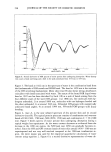
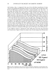
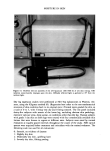
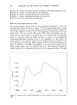
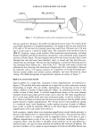
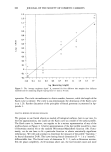
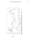
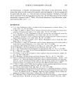
156 JOURNAL OF THE SOCIETY OF COSMETIC CHEMISTS t. 400' 200' t. 000' 800' 600' ß 400' ß 200' 0.000- CH NH C=• CH -.200 ' i ' I ' I ' ! ' ! i I t200 t400 t600 IS00 2000 2200 2400 NAVELENGTH (hi) Figure 4. NIR spectrum of desiccated porcine skin (A) and its second derivative (B). Packard 1000 microcomputer. Diffuse reflectance spectra were collected and trans- formed to absorbance (log l/R) using a 4-nm stepsize over the wavelength range of 1100 nm to 2500 nm. Ratioing of the sample reflectance to the reflectance from the walls of the integrating sphere occured at each wavelength before indexing to the next wavelength. The Technicon solid sampling drawer was customized to accommodate a temperature-stabilized circulating water bath (Figure 1) so that experiments could be conducted at body temperature. The standard Technicon sampling cup was customized with the ceramic insert extending almost to the level of the cover in order to hold the skin taut. The sample cup cover was used with or without a quartz window, depending on the nature of the experiment (Figure 2). This design resulted in a transflectance experiment, i.e., a combination of transmission and reflectance so that the optical pathlength was two to three times the thickness of the skin. Regression analysis was performed with the Technicon standard software COMBO (all-possible combination wavelength search). Sterile porcine skin (Genetic Labs) was used for all in vitro testing. Samples were kept frozen until ready for use. When thawed, the 3" X 12" porcine skin samples were cut into 4-cm circles with a cutting die and kept in a desiccator until treated. The skin samples were hydrated in a closed 96% humidity jar for 24 hours. A sample was re- moved from the jar, weighed on an analytical balance, and placed into the sealed sam- pling cup with quartz window to prevent further evaporation. Then the sampling cup was placed into the 37øC sampling drawer and scanned with the near-infrared spectro- photometer from 1100 to 2500 nm. After the first measurement, the sample was re-

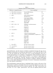
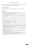
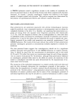
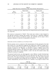
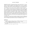
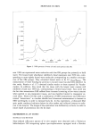
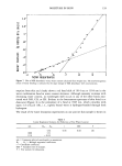
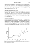
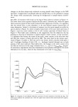
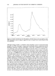
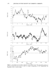
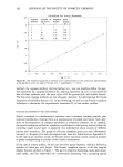
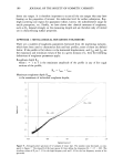
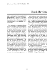
MOISTURE IN SKIN 157 t. 600' i.400' 1.200' z • 1.000- ß RO0. 600 400 1200 i400 1600 1800 2000 WAVELENGTH (nm) 2200 2400 Figure 5. NIR spectra of moist porcine skin undergoing natural water desorption during a weight loss experiment. Water desorption causes a decrease in absorbance of the band around 1900 nm. moved from the sampling cup and allowed to desorb water into the environment or in a dessicator layered with Drierire ©. The drying, weighing, and scanning cycle was con- tinued until a constant dry weight was obtained. Six replicate experiments were com- bined into a single data file that included a total of 50 weight loss measurements. Optical spectra for each weight loss was acquired in triplicate by rotating the sample cup for a total of 150 spectra. Skin samples were also hydrated by treating with solutions of various concentrations of glycerol in water. The samples were scanned in the NIR following equilibration at 40% relative humidity for twenty-four hours. Samples were run in triplicate by rotating the sample cup. Water desorption kinetic studies on the porcine skin were conducted by hydrating the skin in the manner described above. The samples were placed into a windowless sam- pling cup and inserted into the 37øC sampling drawer. NIR spectra were taken every two minutes over the course of eight hours. Kinetic runs were also performed on glyc- erol/water- and skin lotion-treated skin. RESULTS AND DISCUSSION (IN VITRO) UNTREATED PORCINE SKIN-DESORPTION/WEIGHT LOSS The near-infrared spectra of water (trace A) and of moist skin (trace B) are shown in
Purchased for the exclusive use of nofirst nolast (unknown)
From: SCC Media Library & Resource Center (library.scconline.org)