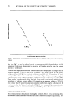
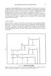
86 JOURNAL OF THE SOCIETY OF COSMETIC CHEMISTS somes. Liposomes are often included in cosmetics to hydrate skin and have been used topically to deliver drugs intercellularly (4). We recently described a method to deliver intracellularly encapsulated purified recom- binant T4 endonuclease V (endo V), a DNA repair enzyme specific for UV-induced pyrimidine dimers (5). Endo V initiates DNA repair by producing a single-stranded break in DNA at the site of damage. We described here our results using endo V liposomes on human epidermal keratinocytes and mouse skin in vivo. MATERIALS AND METHODS The preparation of liposomes containing T4 endonuclease V has been described (5), and the encapsulation efficiency was between 1 and 10%. The liposomes, here designated T4N5 liposomes (patent pending), were composed of phosphatidylcholine, phosphati- dylethanolamine, oleic acid, and cholesteryl hemisuccinate in a 2:2:1:5 molar ratio. The assay for encapsulated enzyme activity has been described (5), and the activity of encapsulated endo V was confirmed in each batch of liposomes. The assay for the pH sensitivity of liposomes is based on the quenching of the fluorescent probe 8-amino- naphthalene-l,3,-6-trisulfonic acid (ANTS) by a high concentration of p-xylene-bis- pyridinium bromide (DPX) entrappeal within the liposome (6). Leakage from the lipo- some diluted DPX relative to ANTS, the quenching was reduced, and fluorescence was increased, as measured with a Hoefer TK-100 fluorometer. All measurements were made in duplicate and averaged. Normal human epidermal keratinocytes and keratinocyte growth media were from Clonetics Corporation (San Diego), and all cell culturing was performed with antibi- otics under sterile conditions without microbial contamination. Unscheduled DNA synthesis (UDS) was measured by growing keratinocytes on glass coverslips and irra- diating them with either 10 or 25 J/m 2 or UV-C from a Philips G15T8 germicidal bulb, monitored by a UVX digital radiometer and UV-C probe. The cells were incu- bated with T4N5 liposomes and 10 •Ci/ml 3H-thymidine (60 Ci/mmol). After four hours the cells were chased with 0.1 mM cold thymidine for one hour, fixed, and coated with Kodak NTB nuclear track emulsion. After seven days the slips were developed with Kodak D-19 developer, and for each treatment grains over the nuclei of 25 lightly labeled cells were counted microscopically and averaged. Loss of endonuclease-sensitive sites (ESS) was measured by growing keratinocytes in 10-cm dishes, irradiating them with 20 J/m 2 of UV-C, and incubating them for 24 hours with media containing T4N5 liposomes. The DNA was purified from cells and treated either with or without T4 endonuclease V, which produced single-stranded breaks at the site of pyrimidine dimers. The average length of the DNA was determined by alkaline agarose gel electrophoresis, and the frequency of ESS, i.e. pyrimidine dimers, was calculated by comparing endo V treated and untreated samples (7). All ESS measurements were performed at least in duplicate and the data averaged. Six-to-eight-week-old female SKH-1 hairless mice (Charles River Labs) were irradiated unrestrained from above with UV-B from two FS40 bulbs monitored with the UVX radiometer and UV-B probe, and each mouse was treated with 0.25 g of T4N5 lipo- somes dispersed in Johnson's baby lotion immediately before use. After six hours the mice were sacrifled and the DNA extracted from epidermis directly over the spine. The

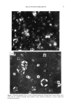
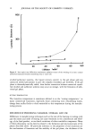
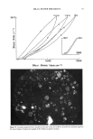
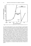
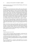
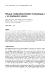
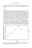
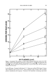
DNA REPAIR IN SKIN 87 frequency of pyrimidine dimers in DNA was measured by the alkaline agarose gel assay described above. All measurements were performed at least in duplicate and the data averaged. RESULTS Liposome-mediated delivery of encapsulated drugs proceeds either by membrane fusion or by endocytosis (8). We have sought to maximize these interactions by constructing liposome membranes that (a) resemble keratinocytes in high phosphatidylcholine con- tent to enhance fusion and (b) are destabilized by the low pH found in internal cellular compartments to release their contents. To measure the ability of recombinant endo V to withstand release into the pH 5 environment of coated vesicles, the purified enzyme was incubated at acidic pH for one hour before the substrate was added and the re- maining activity was assayed. As shown in Figure 1, endo V lost function as the pH declined but retained at least 25% of its activity after one hour at pH 5. These results probably understate the remaining activity, since incubation at acidic pH followed by assay at neutral pH showed greater enzyme activity (unpublished observations). Liposomes sensitive to acidic pH were prepared by combining phosphatidylethanol- amine with oleic acid and cholesteryl hemisuccinate. These components form lipid bi- layers when negatively charged at neutral pH but fail to stack in bilayers when proton- ated in acidic pH (6). To measure the pH sensitivity, T4N5 liposomes were prepared that encapsulated 12.5 mM ANTS and 45 mM DPX instead of endo V. The liposomes were diluted into a pH 5 or pH 8 buffer, incubated at 37 C, and the fluorescence 100' 80' 60' 4.0' 20' 0 I I I I I I I 5 5.4 5.8 6.2 6.6 7 7.4 7.8 pH Figure 1. Activity of T4 endonuclease V at acidic pH relative to optimal pH. T4 endonuclease V was incubated at various pH for one hour. UV-DNA substrate was added and the remaining activity assayed. Results are plotted relative to activity at optimal pH of 6.6.
Purchased for the exclusive use of nofirst nolast (unknown)
From: SCC Media Library & Resource Center (library.scconline.org)