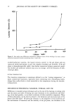
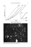
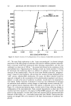
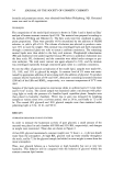
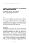
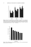
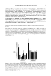
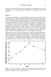
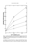
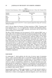
90 JOURNAL OF THE SOCIETY OF COSMETIC CHEMISTS Table I Pyrimidine Dimers Remaining in DNA of UV-Irradiated Keratinocytes 24 Hours After Treatment With T4N5 Liposomes Liposome Dimers/million Percent treatment Ixg/ml bases control None 0 13.7 100 Inactive 0.2 13.7 100 Active 0.05 9.8 71 Active 0.1 7.8 57 Active 0.2 4.7 34 Keratinocytes irradiated with 25 J/m 2 of UV-C. Inactive liposomes prepared by boiling endo V and encap- sulating in liposomes. endo V did not change the frequency of dimers remaining in DNA. Treatment with active liposomes at 0.5, 1.0, or 2.0 •xg/ml endo V showed a dose-dependent enhance- ment of dimer removal. At the highest liposome concentration only one third of the dimers remained as in untreated cells. The effect of T4N5 liposomes in vivo was measured in UV-B-irradiated SKH-1 mice, a strain widely used as a model for human skin in photocarcinogenesis studies. Six-week- old female mice were irradiated with UV-B and treated with liposomes, either immedi- ately after, repeatedly after, or immediately before irradiation (Table II). The dimer frequency in epidermal DNA was compared between mice treated with active or inac- tive liposomes. Mice treated with active liposomes immediately after irradiation had fewer dimers than mice treated with inactive liposomes at each concentration. An op- timal concentration of 0.5 mg/ml endo V in liposomes maximized repair, and higher concentrations did not increase dimer removal. Repeat treatment of mice three hours after the initial application did not further increase repair. Treatment of mice with 2 •xg/ml active liposomes immediately before irradiation was as effective in enhancing repair as was posttreatment. Treatment of mouse skin with inactive liposomes and unencapsulated endo V produced no effect (data not shown), indicating the importance of liposome encapsulation. DISCUSSION T4 endonuclease V, which performs the first step of excision repair of pyrimidine dimers, is able to stimulate DNA repair in cultured XP cells when added by the labora- tory methods of cell permeabilization, microinjection, or DNA transfection (reviewed in reference 11). We have developed a practical method to deliver endo V to cells in culture or in skin by encapsulation in liposomes (5). The lipid membrane of the lipo- somes matched the composition of keratinocyte cell membrane and was destabilized by low pH present in the cell interior. Addition of these liposomes containing endo V to UV-irradiated normal human keratinocytes stimulated DNA repair synthesis (Figure 3) and enhanced dimer removal (Table I). Liposome-based delivery of DNA repair enzymes is a novel and practical method to enhance DNA repair in skin. The SKH-! hairless mouse is often used as a model for human skin in studies on photoaging and photocarcinogenesis, and its skin structure

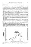
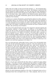
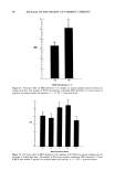
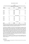
DNA REPAIR IN SKIN 91 Table II Pyrimidine Dimers Remaining in Epidermal DNA of UV-B-Irradiated SKH-1 Mice 6 Hours After Treatment With T4N5 Liposomes Liposome UV-B Dimers per Percent treatment •g/ml J/m 2 million bases control Posttreatment Inactive 0.1 10,000 92.9 100 Active 0.1 10,000 55.9 60 Inactive 0.5 10,000 90.8 100 Active 0.5 10,000 23.8 26 Inactive 2.0 10,000 93.0 100 Active 2.0 10,000 72.9 78 Repeat application Inactive 0.1 2,500 43.7 100 Active 0.1 2,500 22.0 50 Inactive 1.0 2,500 36.4 100 Active 1.0 2,500 23.8 65 Inactive 2.0 2,500 39.8 100 Active 2.0 2,500 26.8 67 Pretreatment Inactive 2.0 10,000 79.0 100 Active 2.0 10,000 63.6 80 Posttreatment: liposomes added immediately after UV-B irradiation. Repeat application: liposomes added immediately after and again three hours after UV-B irradiation. Pretreatment: liposomes added immedi- ately before UV-B irradiation. Inactive liposomes prepared with boiled endo V. and thickness is comparable to human skin in the temple region of the forehead. T4N5 liposomes reduced the frequency of pyrimidine dimers in SKH-1 mouse skin by about 50% when applied either immediately after or immediately before UV irradiation (Table II). Repeated application of the liposomes did not further enhance repair, sug- gesting that repair was not limited by the loss of endo V activity in the initial liposome treatment. These results suggest that the liposomes penetrate the stratum corneum of the skin and enter keratinocytes. Keratinocytes in culture showed enhancement of re- pair with increased liposome concentration, while mouse skin showed a non-linear re- sponse, with a concentration optimum. These differences may reflect the complexity of liposome penetration and enzyme uptake. Our goal is for T4N5 liposomes to be included in either sunscreen formulations for use before sunning or in cosmetic preparations for regular use to repair DNA damage from exposure to solar UV. REFERENCES (1) A. Glass and R. Hoover, The emerging epidemic ofmelanoma and squamous cell skin cancer,JAMA, 262, 2097-2100 (1989).
Purchased for the exclusive use of nofirst nolast (unknown)
From: SCC Media Library & Resource Center (library.scconline.org)