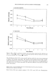
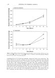
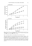
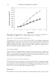
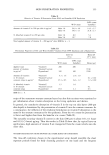
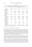
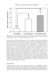
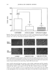
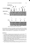
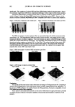
150 JOURNAL OF COSMETIC SCIENCE components with an Ikavisc MR-D1 mixer (Janke & Kunkel) for 30 minutes during a heating and cooling cycle between 25 øC and 95 øC. The emulsions were homogenized for five minutes at 40øC. The w/o cream was made by heating the oil/emulsifier mixture to 80øC and stirring in the hot water phase. The mixture was stirred for five minutes at 80øC and then cooled to room temperature while being stirred. The shower gel was prepared by mixing the components for 30 minutes at room temperature. Phase behavior of the emu/siom. Liquid crystalline phases were identified by polarization microscopy (Zeiss, K61n, Germany). The emulsion type (o/w or w/o) was determined by conductivity measurements (Radiometer, Copenhagen, Denmark). Viscosity measureme, t. Flow and viscosity curves in the shear rate range from 0 to 100/s were studied with a thermosrated, shear-rate-controlled rotation rheometer RFS 2 (Rheometrics, Piscataway, NJ) with a plate-plate measuring system (2 mm gap) at 25 øC. Partide sizes. The particle size distribution in the undiluted emulsions was determined with an optical microscope with the help of the Optimetrix digital image analyser (Stemmer, Meerbusch, Germany). Bovine udder skin (B US) model. The in vitro "isolated perfused bovine udder skin" model makes use of material from slaughterhouses. Immediately after an animal has been slaughtered, the udder skin is perfused with heated and oxygen-enriched Tyrode's solution (80-100 mm Hg pressure approx. 120 ml/minute) under laboratory condi- tions. The viability of the skin is monitored biochemically in the perfusate by deter- mining the pH, the lactate-dehydrogenase activity, and the lactate and glucose concen- trations. The skin surface temperature (approx. 32øC) and skin fold thickness 3mm are measured physically (9). The skin of the udder is thin and exhibits all the morphological characteristics of mammalian skin, including the cutaneous appendages such as seba- ceous glands and hair follicles. The skin of the udder functionally resembles human skin (9). The experimental procedures such as preparation, perfusion, viability checks, the topical application of the coded test substances, and sampling were performed at SIMRED GmbH (Gro[3burgwedel, Germany). Skin penetration. The penetration of oil- and water-soluble vitamins from the different formulations into perfused bovine udder skin was studied in two ways. Leave-on application. Fifteen minutes after perfusion started, 3 g of each different vita- min-containing cream formulation were applied topically to skin areas measuring 75 cm 2. This high dosage was intended to prevent any depletion of the vitamin concen- tration in the vehicle during the penetration experiment (infinite dose). After one hour and five hours, respectively, the residual cream was carefully removed with a paper towel, and then adhesive tape strips and samples for preparing dermatome sections were taken. The adhesive strips and skin samples were kept deep-frozen at -20øC. Rinse-off application. Fifteen minutes after perfusion started, 3 g of each different vi- tamin-containing shower gel formulation were applied topically to skin areas measuring 75 cm 2. After two minutes the shower gel was rinsed off with a surplus of warm water (40øC). The skin was carefully dried with a paper towel. This washing-rinsing procedure was repeated three times, and then adhesive tape strips were taken.

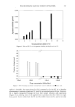
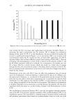
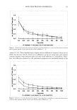
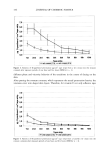
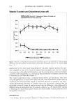
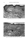
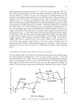
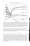
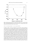
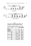
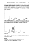
SKIN PENETRATION PROPERTIES 151 Adhesive tape stripping. Stripping (Tesa © 4204, BDF, Hamburg) was used to remove the outermost layers of the stratum corneum in sequence about ten layers of horny-layer cells (roughly 10 l•m) were removed. The mass of the removed horny-layer cells was constant for all layers, being approx. 1.10 mg per adhesive strip (1.9'10 cm), with a standard deviation of +0.36 mg (n = 20). The dermatome sections parallel to the skin surface were cut into 20-pm-thin sections and went down 200 pm through the epider- mis into the upper layer of the dermis. Detection of the vitamins. The detection of vitamin E, vitamin E acetate, and D-panthenol in the adhesive strips from the outermost skin layer and in the dermatome sections of the dermis and epidermis was based on extraction of the analytes and subsequent liquid chromatographic analysis of the obtained extracts. The selective determination of the vitamins in the complex matrix required substance-specific detection to be carried out after the chromatographic separation. The quantification was based on external standard calibrations and comparison of the treated and untreated skin. Detection of vitamin E and vitamin E acetate. Vitamin E/vitamin E acetate were extracted from the relevant skin sections with ethanol, and then the solvent was quantitatively removed and the residue was taken up in hexane. Extraction from adhesive strips was carried out directly with hexane. Vitamin E was separated from unwanted accompanying substances in a diol phase (Lichrospher 100 Diol) by means of liquid chromatography. The chromatography was carried out under isocratic conditions with n-hexane/te•-t. butyl methyl ether as eluent. Selective detection was performed with a fluorescence detector. The chromophoric system in the vitamin E molecule enabled the work to be carried out at an excitation wavelength of 295 nm. Detection ofpanthenol. In contrast to vitamin E and vitamin E acetate, panthenol has no molecular properties that would facilitate sensitive and selective detection in difficult matrices. After extraction from the skin or adhesive strips, therefore, the obtained extract was first hydrolyzed alcoholically and the panthenol quantitatively converted to ami- nopropanol. This hydrolysis product was separated from unwanted accompanying sub- stances in an ion-exchange column with diluted sodium hydroxide solution as eluent, and was converted to a strongly fiuorescing isoindole derivative in a postcolumn reaction with orthophthaldialdehyde. The fluorescence emission was measured with an HPLC fluorescence detector at a wavelength of 455 nm. RESULTS AND DISCUSSION PHASE BEHAVIOR OF THE COSMETIC FORMULATIONS For the leave-on products, consideration also has to be given to their behavior after open topical application. When they are applied, the emulsions are spread over the skin, forming a film (approx. 400 l•m). As the temperature of the emulsions increases to skin temperature, components with a high vapor pressure, in our case water, start to evaporate (3,4). Drying experiments on thin emulsion films show that•clepending on atmo- spheric humidity and the thickness of the layer--almost all the water escapes from the emulsions within five to ten minutes. During this period the viscosity and sometimes also the structure of the emulsion change (10).
Purchased for the exclusive use of nofirst nolast (unknown)
From: SCC Media Library & Resource Center (library.scconline.org)