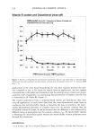
j. Cosmet. sci., 50, 159-170 (May/June 1999) Histopathological, morphometric, and stereologic studies of dermocosmetic skin formulations containing vitamin A and/or glycolic acid PATRICIA MARIA BERARDO GON(•ALVES MAIA CAMPOS, GISLAINE RICCI, MARISA SEMPRINI, and RUBERVAL A. LOPES, Faculty of Pharmaceutical Sciences of Ribeira7o Preto (P.M. B. G. M.C., G.R.) and Dental School of Ribeir•o Preto (M.S., R.A.L.), University of Sa7o Paulo, Sa7o Paulo, Brazil. Accepted for publication April 3 O, 1999. Synopsis Among the many active agents for dermocosmetic purposes that have been described, marketed, and prescribed, vitamins (vitamin A palmitate among them) and alpha-hydroxy acids such as glycolic acid have been gaining scientific importance. Vitamin A palmitate contributes to the maintenance of skin softness and smoothness, improving the water barrier properties of the tissue. Glycolic acid has yielded highly satisfac- tory results in terms of recovery of aged skin. The combination of low concentrations of glycolic acid with vitamin A palmitate has been extensively used in dermocosmetic formulations. The objective of the present study was to investigate the histopathological alterations caused by formulations containing vitamin A and/or glycolic acid in guinea pig skin, determined by appropriate stereologic techniques. The following formulations were applied to specific shaved areas of guinea pig skin: gel alone (used as the dermocosmetic base), gel with vitamin A added, gel with glycolic acid added, and gel with both vitamin A and glycolic acid added. After application of the formulations for one week, skin biopsies were obtained from the animals and we investigated the histopathological alterations. Under the present experimental conditions, both the formulations containing vitamin A and glycolic acid caused increased epidermal thickness, with cells of larger volume due to intra- and extracellular edema (hydration). This epithelial thickening was not limited to the upper cell layers but was also present in the basal and spinous layers. These alterations were even more evident with the use of the formulation con- taining a combination of vitamin A and glycolic acid. INTRODUCTION Among the countless active principles used for dermocosmetic purposes, vitamins (and vitamin A palmitate among them) and alpha-hydroxy acids (such as glycolic acid) have gained notoriety due to their pharmacodynamic properties (1). Alpha-hydroxy acids, a group of natural substances found in fruits and in other foods, have been widely used by the cosmetics industry for the preparation of anti-aging skin formulations. 159

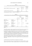
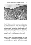
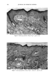
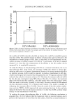
160 JOURNAL OF COSMETIC SCIENCE Vitamin A palmitate acts on the skin by keeping it in good condition and favoring its correct metabolism. It acts on epithelization in dry and rough skin, as well as on keratinization considered to be abnormal. Vitamin A palmirate has been used in der- mocosmetic preparations in combination with glycolic acid because the latter reduces the cohesion of corneocytes, stimulating skin desquamation, which in turn facilitates vitamin A absorption by the skin and leads to the expected results. Alpha-hydroxy acids are being incorporated into a new generation of treatment cosmet- ics. This is a new and interesting chapter in the formulation and scientific investigation of these products, since satisfactory results in the recovery of aged skin can be obtained with much simpler molecules. The objective of the present study was to investigate the histopathological alterations caused by dermocosmetic formulations containing vitamin A and/or glycolic acid in guinea pig skin, determined by appropriate stereologic techniques. EXPERIMENTAL PROCEDURE FORMULATIONS STUDIED We used a non-ionic gel formulation consisting of 2% hydroxy-ethyl-cellulose, 2% glycerin, 3% propyleneglycol, 0.2% methyldibromo-glutaronitrile and phenoxyethanol, 0.01% alpha-tocopherol and distilled water, with and without the addition of 0.5% vitamin A palmirate (1,000,000 IU/g) and/or 4.2% glycolic acid. BIOLOGICAL ASSAY Artfinals. Adult guinea pigs weighing on average 350 g were used. The animals were kept in individual cages and received commercial ration and green food (rami), as well as water ad/RSftum. Treatment. Five areas measuring 1 cm in diameter were shaved on each side of the dorsum of each animal, one of them used as control and the other four for the application of the cosmetic formulations. The formulations were applied daily for one week. The treatment was as follows: a) area I, no treatment (control) b) area II, application of the gel only c) area III, application of the gel with vitamin A added d) area IV, application of the gel with glycolic acid added e) area V, application of the gel with vitamin A and glycolic acid added. Histology. After one week of treatment the guinea pigs were sacrificed and skin fragments were obtained from each shaved area and immediately immersed in a fixing solution of 85 ml of 80% alcohol, 10 ml formalin, and 5 ml acetic acid. The fragments were fixed for 24 hours and then dehydrated, cleared, and embedded in paraffin. Serial 6-pm-thick sections were then obtained, and ten sections per block were obtained from a total of 500 sections, so that each of these ten sections would correspond to an interval of 50 sections. The sections were stained with hematoxylin and eosin. 3•orphometry and/•aryometry. For the morphometric study (analysis of the nucleus of the epithelial layers), the skin sections obtained from each experimental group were analyzed with a Henamed light microscope equipped with a 100x immersion objective and a light camera (Jena).
Purchased for the exclusive use of nofirst nolast (unknown)
From: SCC Media Library & Resource Center (library.scconline.org)