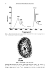
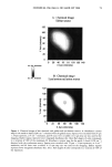
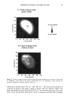
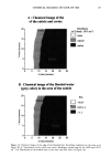
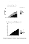
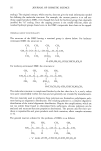
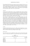
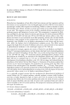
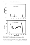
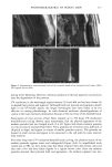
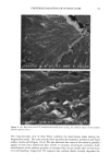
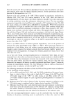
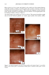
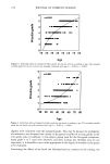
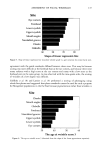
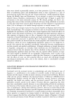
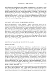
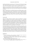
88 JOURNAL OF COSMETIC SCIENCE 15- P 10- ._ E ._ - 5 A: Chemical Image of the of the cuticle and cortex 5 10 15 20 X Axis (microns) Absorbance (vCH band' 2875 cm 4) D resin cuticle I cortex B ß Chemical image of the Bonded water (grey color) in the area of the cuticle 15. P lO ._ E x - 5 /. .. B/A • 0.85 I 0.85-1.1 1.1 0 5 10 15 20 X Axis (microns) Figure 13. Chemical images of the edge of untreated hair. Recording conditions are the same as in Figure 5B and Figure 12. A: Visualization of the cuticle and cortex: absorbance contour map for the vCH band (2875 cm ]). B: Distribution of the bonded water in the same area (B/A ratio: see Figure 10).

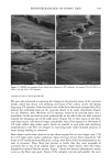
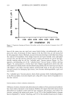
CHEMICAL IMAGING OF HAIR BY IMS 89 reveals that the bonded water is present everywhere in the cuticle but not in the cortex. The same kind of images have been obtained on the untreated hair (Figure 13A,B). In this case, there is no water localized in the cuticle. The experiments reveal, therefore, the presence of bonded water within the cuticle, correlated to the bleaching process. Once again, several different samples were measured with similar findings. The results suggest a structural alteration of the cuticle in the bleached hair. The oxidative scission of the disulfide bonds induces a decrease in the density of the crosslinked protein and provides anionic sites in the form of cysteic acid residues (2). This leads to an enhancement of the hydrophilicity of the fiber and contributes to the increase in swelling (2). In addition, moisture absorption increases with the extent of bleaching from 13.5 % for intact hair to 14.8% for bleached hair (2). Therefore, the preferential water affinity of the cuticle could be explained by a preferential hydrophilicity and moisture absorption of this cuticle induced by the bleaching. CONCLUSION Using synchrotron infrared microspectroscopy, details in a chemical imaging of a hair cross section with very high contrast are shown for the first time. The cortex can easily be differentiated from the cuticle and the medulla. In addition, details in the areas of the cuticle and the medulla are shown with a lateral resolution of 4 microns. The effect of hair bleaching has also been studied, and we point out the presence of bonded water within the cuticle. In particular, we have studied the spatial distribution of the sulpho- nate contributions, which show non-homogeneous distribution within the hair. There- fore, the combination of IR microspectroscopy and synchrotron radiation is a powerful tool for the spatial and chemical resolution of hair regions of only a few microns. This opens up chemical imaging characterization of the interaction of hair with chemical reagents at resolutions of a few microns. ACKNOWLEDGMENTS We are very grateful to Paul Dumas for his assistance and very fruitful discussions and to Jean-Marc Mouchon and Marc Piscaglia for the material preparation. The Irus mi- crospectrometer was provided by the Northrop Grumman Corporation as part of a collaborative research program with the NSLS. The results presented in this work are part of an ongoing study supported by the Elf Aquitaine company. The National Synchrotron Light Source is supported by the United States Department of Energy under contract DE-AC02-98CH 10886. REFERENCES (1) E.G. Bendit, Infrared spectrum of keratin. I. Spectra of or-, [3-, and supercontracted keratin, Biopol- ymers, 4, 539-559 (1966). (2) J. Jachowicz, Hair damage and attempts at its repair, J. Soc. Cosmet. Chem., 38, 263-286 (1987). (3) E. Hoting, M. Zimmermann, and H. Hocker, Photochemical alterations in human hair. Part II: Analysis of melanin, J. Soc. Cosmet. Chem., 46, 181-190 (1995). (4) J. Strassburber, Quantitative Fourier transform infrared spectroscopy of oxidized hair, J. Soc Cosmet. Chem., 36, 61-74 (1985).
Purchased for the exclusive use of nofirst nolast (unknown)
From: SCC Media Library & Resource Center (library.scconline.org)