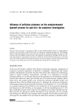
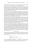
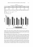
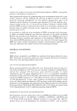
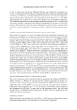
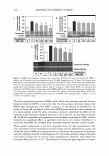
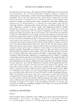
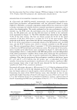
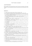
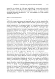
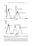
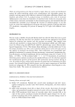
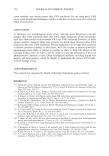
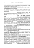
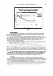
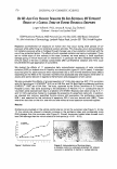
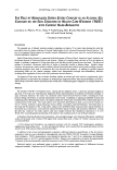
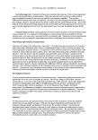
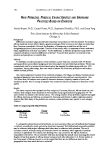
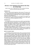
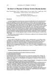
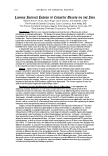
(a) (b) � = Q u � = Q u DELIVERY ACTIVITY OF pH-SENSITIVE LIPOSOMES 147 PC:CHEMS liposome 10• FU-Height w• FU-Height Figure 4. Histograms of cellular fluorescent intensity of HM3KO cells treated with PC:CHEMS and DOPE:CHEMS liposomes containing 1 % fluorescent-DHPE (a) and treated with calcein-loaded DOPE: CHEMS and PC:CHEMS liposomes (b) as measured by FACS. ery and the internalization mediated by pH-sensitive DOPE:CHEMS liposomes. DOPE: fluorescein-DHPE:PEG-5 rapeseed sterol (L5) liposome was used as a control for pH- insensitive liposomes. Cellular uptake behavior of the pH-sensitive liposomes was moni- tored by using 1 % fluorescein-DHPE incorporated in the liposomes. Simultaneously,

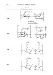
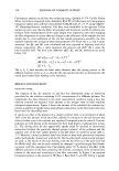
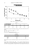
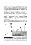
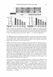
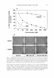
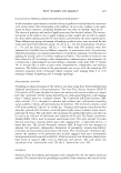
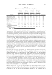
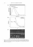
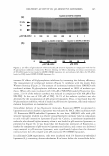
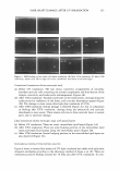
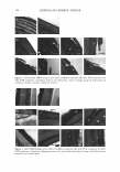
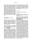
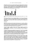
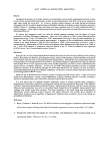
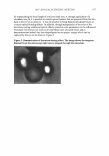
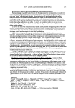
148 JOURNAL OF COSMETIC SCIENCE the release of molecules loaded in the pH-sensitive liposomes was evaluated by encap- sulating dextran-rhodamine B in the liposomes. Figure 5 was obtained from a repre- sentative CLSM experiment following one hour of incubation of HM3KO cells with 1 % fluorescein-embedded and dextran-rhodamin-B-loaded pH-sensitive and pH-insensitive liposomes. Fluorescent intensity from fluorescein and rhodamine B is significantly high in cells incubated with pH-sensitive liposomes (L) as compared with the control. The results of calcein and dextran-rhodamin B delivery by pH-sensitive liposomes correlate Figure 5. CLSM images of HM3KO cells after incubation with DOPE:PEG-5 rapeseed sterol (L5) (a,b,c) and DOPE:CHEMS (L3 ) (d,e,f) liposomes containing 1 % fluorescent-DHPE at 37 ° C for one hour. Cells are under a fluorescein filter (a and cl) and under a rhodamine filter (b and e). Part c is a composite of superimposed layers from a and b and an image under a DAPI filter. Part f is a composite of superimposed layers from cl and e and an image under a DAPI filter. Scale bar = 20 µm.
Purchased for the exclusive use of nofirst nolast (unknown)
From: SCC Media Library & Resource Center (library.scconline.org)