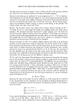
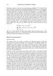
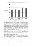
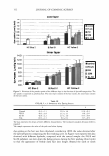
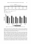
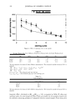
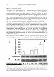
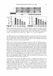
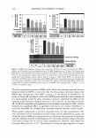
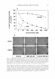
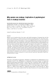
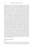
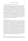
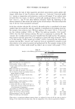
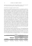
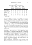
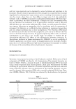
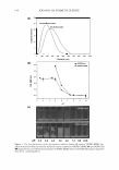
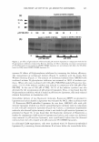
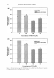
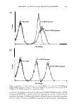
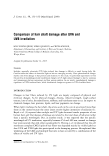
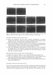
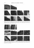
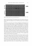
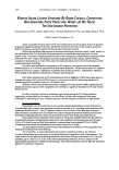
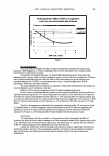
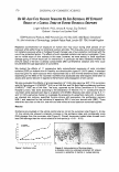
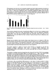
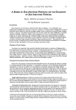
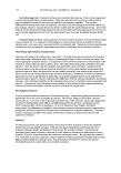
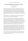
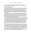
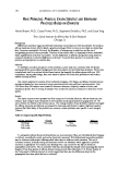
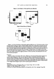
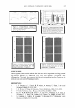
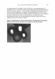
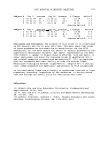
HAIR SHAFT DAMAGE AFTER UV IRRADIATION 153 Figure 1. SEM findings in hair shafts: (A) Before irradiation. (B) After UVA irradiation. (C) After UVB irradiation. Arrow: focal lifts at edges of cuticles. Arrowhead: focal losses of curicular edges. Conventional transmission electron microscopic study (a) Before UV irradiation: The hair shows concentric arrangements of smoothly bounded cuticular cells containing the normal complement and distribution of the A-layer, exocuticle, and endocuticle subcomponents (Figure 2A). (b) After UV A irradiation: Variable-sized holes in the endocuticles, cleavage along the endocuticles by confluence of the holes, and cuticular detachment appear (Figure 2B). The damage is more severe after high-dose irradiation of UVA. (c) After UVB irradiation: Similar damage is observed (Figure 2C), but in comparison to findings after UV A irradiation, cleavage along the endocuticle and cuticular detachment is more severe. There remain only two to three cuticular layers in several parts, due to extensive damage. Lipid transmission electron microscopic study with special fixative (a) Before UV irradiation: There are intact intercellular lipid layers (Figure 3A). (b) After UVA irradiation: There are some bulging portions in the intercellular lipid layers and small focal lacunae along the intercellular spaces (Figure 3B). (c) After UVB irradiation: Several bulging portions in the intercellular lipid layers are also observed (Figure 3C). BIOCHEMICAL FINDINGS WITH PROTEIN ANALYSIS Figure 4 shows a western blot analysis of UV-light-irradiated hair shafts with polyclonal ubiquitin antibodies according to the laboratory method of Inoue et al. (8). There are continuous positive findings around the 10 kDa area after UV A irradiation. In com-

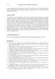
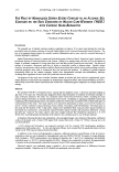
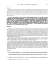
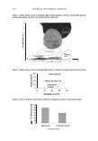
154 JOURNAL OF COSMETIC SCIENCE Figure 2. Conventional TEM findings in hair shafts: (A) Before irradiation. (B) After UVA irradiation. (C) After UVB irradiation. Arrowhead: holes in the endocuticles. Arrow: cleavage along the endocuticles by confluence of holes. Asterisk: confluence of holes. Figure 3. Lipid TEM findings in hair shafts: (A) Before irradiation. (B) After UV A irradiation. (C) After UVB irradiation. Arrowhead: bulging portions in the intercellular lipid layers. Arrow: small focal lacunae along the inrercellular spaces.
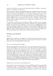
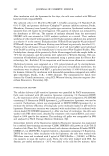
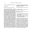
Purchased for the exclusive use of nofirst nolast (unknown)
From: SCC Media Library & Resource Center (library.scconline.org)