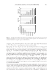
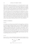
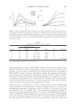
EFFECT OF Gp4G ON SKIN TISSUES 471 applications, including anti-aging and skin repair (16,17). Although Gp4G has been on the market for several years, its action mechanisms are unknown. Moreover, there is no published data showing how Gp4G works on skin components and its effects on the fol- licle cycle. Therefore, the aim of this work is: (i) to analyze whether the Gp4G formulation stimulates hair growth (ii), to characterize morphophysiological parameters in the der- mal tissues, and (iii) to propose a biochemical mechanism for its effect. MATERIALS AND METHODS Gp4G FORMULATION Gp4G was extracted from CAS by FarmaService Bioextract in São Paulo, Brazil, using a traditional method (14), and its concentration was checked by an adapted capillary elec- trophoresis method (18,19). Gp4G extracted from CAS stock solution was used either as a pure solution prepared in MilliQ-water in cell culture studies or as a Gp4G formulation to enhance skin penetration in in vivo experiments, which consisted of a phospholipid vesicle suspension supplemented with calcium and red pepper extract containing 0.69 g/l of gallic acid equivalents in total polyphenols and 1.70 g/l of Trolox equivalent in an antioxidant capacity (DPPH suppression) measured according to the specifi ed literature (18–21). Soybean lecithin was dissolved in a volatile organic solvent, which was evaporated under argon fl ux and rehydrated with Gp4G stock solution and calcium chloride 1.0 mM, and fi nally sonicated for fi ve minutes. The fi nal concentration of lecithin was 2 mg/ml. A control group containing only Gp4G and liposomes was also implemented, showing results similar to those reported for the Gp4G formulation, but in smaller magnitude. We suspect that this is due to the fact that both red pepper and calcium are important in allowing better penetration of Gp4G in the skin. IN VIVO HAIR ELONGATION EVALUATION The Gp4G formulation was topically applied to the dorsal region (8 cm2, shaved) of Wistar rats (n = 5). The control group was not treated with the product (n = 5). Male rats were born and kept in the Animal Care Facility of the Chemistry Institute of the Univer- sity of São Paulo. All rats were of the same age (20 days). Therefore, their follicles were in the fi rst telogen phase, and experiments were followed during a hair follicle cycle induced by depilation (22). About 1 ml of the product was applied daily with a cotton swab over a period of 28 days. At the end of the application period, a sample of hair shaft from the treated skin was collected to measure its width. Hair shafts were measured for each ani- mal using a calibrated ruler under a microscope. The distribution of overcoat and under- coat hair shafts was about 70% and 30%, respectively. These two classes were considered together to calculate a general average size and separately to determine the average length of the independent populations of the hair shafts. The experiment was repeated twice. HISTOLOGICAL STUDIES Results were analyzed qualitatively and quantitatively after morphometric evaluation in comparison with the control group. At the end of the application period the rats were anesthetized to obtain dorsal skin samples that were immediately fi xed properly for his- tological and immunohistochemical analyses. National standards for the care and use of

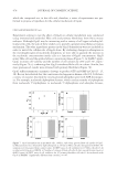
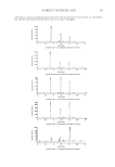
JOURNAL OF COSMETIC SCIENCE 472 laboratory animals, compatible with NIH standards, were followed. All studies were ap- proved by the Institutional Animal Care and Use Committee. LIGHT MICROSCOPY PROCESSING Samples were fi xed for three hours in Methacarn (methanol chloroform glacial acetic acid 6:3:1), rinsed with absolute ethanol, and embedded in Paraplast (Oxford, St. Louis, MO) at 60°C. Tissues were cut on a microtome (Micron HM-200) into 5-μm sections, affi xed to glass slides using 0.1% poly-L-lysine (Sigma, St. Louis, MO), and then dried at room temperature. For morphological analysis, all sections were stained with hematoxylin- eosin. Some sections were histochemically stained by the picrosirius method for evalua- tion of collagen fi ber content and distribution. The observations were performed in a minimum of three sections. ANAGEN/TELOGEN PHASE DISTINCTION The phases of the hair cycle were analyzed at the level of the sebaceous gland. Telogen and anagen hair follicle phases were identifi ed in hematoxylin-eosin–stained histological sec- tions and were classifi ed according to Headington (24). This method provides a simple and reliable way to differentiate hair follicles in anagen and telogen. Accordingly, the medulla of the telogen hair follicles were stained in pink by H&E in the region of the sebaceous glands, and the medulla of anagen hair follicles were not stained in pink at the height of the sebaceous glands (24). IMMUNOPEROXIDASE PROCEDURES Five-micrometer-thick sections were affi xed to glass slides using 0.1% poly-L-lysine (Sigma) and then dried at room temperature. Each of the succeeding steps was followed by thorough rinsing with phosphate-buffered saline (PBS). Sections were treated with 3% H2O2 in PBS for 30 min to block endogenous peroxidase activity. All steps were per- formed in a humidifi ed chamber, and care was taken to avoid the drying out of sections. Antigen retrieval was done by enzymatic treatment of sections before the immunoreac- tion. For versican immunoreaction, sections were incubated (1 h at 37°C) in 20 mM Tris-HCl, pH 6.0, containing 0.2 U/ml of chondroitinase ABC (Seikagaku Corp., Tokyo, Japan). For laminin immunostaining, sections were incubated with a 2 mg/ml solution of porcine pepsin (1,120 units/mg protein) (Sigma) in pH 2.2 acid buffer for 20 min at room tem- perature. Nonspecifi c staining was blocked by incubating the sections (1 hr) with normal goat serum, diluted 1:1 (v/v) in PBS-10% BSA (w/v) (room temperature). The sections were then incubated with rabbit anti-VER polyclonal antibody (Chemicon International, Temecula, CA) diluted 1:500 in PBS-0.3% (v/v) Tween 20 or with rabbit polyclonal antibody against laminin (BioGenex, CA) diluted 1:50 in PBS-0.3% (v/v) Tween 20. After rinsing in PBS, all sections were incubated with biotin-conjugated sec- ondary antibody (goat anti-rabbit IgG) diluted in PBS for one hour at room temperature (25°C). Subsequently, sections were incubated with the streptavidin/peroxidase complex (Vector Laboratories, Burlingame, CA) for one hour at room temperature. Peroxidase was visualized using 0.03% 3,3′-diaminobenzidine in PBS with 0.03% H2O2. To achieve standardization of the immunoreactions for each antibody, the slides from the control and
Purchased for the exclusive use of nofirst nolast (unknown)
From: SCC Media Library & Resource Center (library.scconline.org)