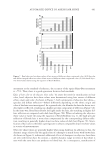
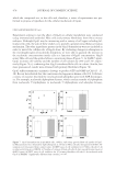
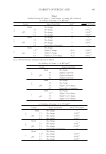
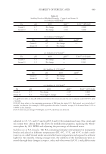
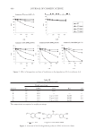
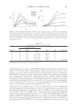
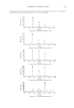
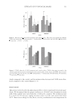
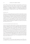
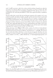
EFFECT OF Gp4G ON SKIN TISSUES 475 facial hair growth since the physiological responses present in human scalp are opposite to those found in facial hair (27). The fact that the Gp4G formulation stimulates follicles suggests that it may also be stimulating adjacent interfollicular and bulge stem cells, which may affect the health of dermal and epidermal tissues (28–30). D. Oh and co-workers (27) have shown that even whole proteins (human growth hormone) encapsulated in liposomes, which are delivered to the hair follicles and allowed to interact with their adjacent stem cells, have shown dramatic and generic effects as hair growth, anti-acne effects, skin tone improvement, and anti-wrinkle effects (27,31). To characterize possible actions of the Gp4G formula- tion on dermal tissues, we have investigated some key morphofunctional parameters such as vascularization, fi broblast activation, and deposition of collagen and proteoglycan versican in the dermis (32). EFFECT ON THE DERMIS Again using Wistar rats that were or were not treated with the Gp4G formulation, tissue samples were taken and evaluated histologically. The number of active fi broblasts (large cells with irregularly branched cytoplasm) was counted in histological hematoxylin- eosin–stained sections of the skin (Figure 3) (26), and it is clearly higher in the Gp4G formulation-treated group compared with the control group. It is expected that a higher number of active fi broblasts leads to increased synthesis and deposition of collagen molecules. In fact, the sections stained using the picrosirius method (33), and analyzed using polar- ized light microscopy, showed a statistically different higher number of thin brilliant green collagen fi brils in skin treated with the Gp4G formulation, whereas in non-treated skin thick yellow brilliant fi brils were predominant (Figure 4). The high proportion of thin fi bers supports our hypothesis that the Gp4G formulation may favor fi broblast acti- vation and de novo synthesis of collagen fi brils in the dermis. Vascularization is another important parameter that can be related to hair growth and the physiological conditions of epidermal tissues. Figure 5 shows sections of skin immunostained with an anti-laminin (LM) antibody. LM is a specifi c marker for basement membranes, present in all types of blood vessels (34,35). Importantly, the number of vessels per unit of area showed an expressive increase (38%) in the group treated with the Gp4G formulation (Figure 5). It has been shown earlier that follicle cycling is associated with Figure 2. Effect of Gp4G formulation on the hair growth phases. Sections of H&E-stained dorsal skin from (a) controls (not treated with Gp4G) and (b) rats treated with Gp4G formulation. (c) Percentage of anagen and telogen hair growth phases. * Signifi cantly different from controls, p 0.001. Arrows indicate the position of the sections in the height of the sebaceous glands.

JOURNAL OF COSMETIC SCIENCE 476 expressive changes in skin perfusion and that the onset of anagen itself is associated with angiogenisis. We have shown that treatment with the Gp4G formulation favors both anagen follicles and angiogenisis. Based on our data, we cannot affi rm whether the effect of angiogesis is a direct effect of the Gp4G formulation or is an indirect effect because of the presence of a higher percentage of anagen follicles. However, the fact that both effects were observed experimentally gives confi dence that our experimentally gives confi dence that our experimental data is self-consistent. Although the data shown in Figures 2–5 gives a strong indication that the Gp4G formu- lation provides direct benefi ts to the dermal tissue, it was important to test the presence of a recognized marker of cellular proliferation. Versican (VER) is a large chondroitin sulfate proteoglycan that belongs to the family of hyaluronan-binding proteoglycans, Figure 3. Effect of Gp4G formulation treatment on the active fi broblasts. H&E-stained paraffi n sections showing the higher number of fi broblasts in the (a) Gp4G-treated skin compared with the (b) non-treated skin. Arrows show examples of active fi broblasts. Figure 4. Picrosirius-stained sections observed under polarized microscopy. Untreated skin contains pre- dominantly red collagen fi bers (a). Thin green and yellow fi bers predominate in Gp4G-treated skin (b). Quantitative evaluation of thin brilliant green and yellow fi bers in a section of Gp4G formulation-treated and untreated skin. Quantifi cations were conducted by elimination of the specifi c color channels (blue and red) after Image J treatment. *Signifi cantly different from controls, p 0.001. Figure 5. Effect of Gp4G formulation treatment on the vascularization of dermal tissues. Average density of subepithelial dermal vessel immunostained with anti-laminin antibody in the (a) Gp4G formulation-treated and (b) untreated skin. (c) Density of vessels/area in skin section in the subepidermal and deep dermis. *Sig- nifi cantly different from controls, p 0.005.
Purchased for the exclusive use of nofirst nolast (unknown)
From: SCC Media Library & Resource Center (library.scconline.org)