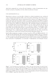
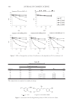
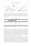
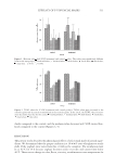
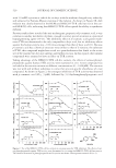
EFFECT OF Gp4G ON SKIN TISSUES 479 promote reversible phosphotransfer between ADP, ATP, and AMP (50). This and other enzymatic systems allow that high-energy phosphates from a triphosphate nucleoside (GTP, for instance) gets transferred to a diphosphate nucleoside (ADP, for instance), which explains the increase in ATP concentration. Quantifi cation of intracellular ATP was conducted using the luciferin/luciferase method (25). In the cells treated with Gp4G, there was a 38% increase in ATP concentration (Figure 8a). This increase in intracellular ATP concentration is equivalent to that observed in Artemia salina (14). Increase in in- tracellular ATP with increases in ATP/ADP ratios are know to trigger several signaling pathways that lead mainly to inhibitory metabolic regulation (respiratory control), which are not compatible with the data on cell viability and tissue activation (51,52). Besides ATP, the asymmetric cleavage of Gp4G and consequent catalyzed phosphotrans- fer systems produces di/monophosphate nucleosides, which are known to regulate several metabolic pathways, including activation of phosphofrutokinase, inhibition of ATPase, stimulation of respiration, and regulation of different types of UCPs (48–55). This could explain the cellular activation that triggers the effect on the dermal tissues shown above. Therefore, it is necessary to quantify if Gp4G treatment would also increase the concen- tration of di/monophosphate nucleosides. Quantifi cation of the intracellular concentra- tion of nucleotides in Hela cells was conducted using HPLC (Figure 8b). Note that the treatment with Gp4G causes an increase in the intracellular concentration of all di/mono- phospate nucleosides, with statistically signifi cant differences for GDP (Figure 8). Paral- lel increases in concentrations of triphosphates and di/monophosphate nucleosides are not common in nature, because these nucleotides are mutually controlled and the increase in ATP concentration usually occurs with the consecutive decrease in the concentrations of ADP and AMP (50). Therefore, the treatment with Gp4G generates a new homeostatic balance in cells, in which the concentration of both triphosphates and di/monophosphate nucleosides are increased. This new equilibrium of tri/di/monophosphate nucleoside concentration may have direct (described above) and indirect effects in cell metabolism. One example of the indirect effect would be the activation of K+ channels, which are known to respond to the intracellular concentration of ADP/GDP (56) that activates tissue vas- cularization. The degree in which particular compounds are able to regulate the opening of K+ channel varies with the tissue type. The relationship between the opening of K+ channels and hair growth has been evidenced as the main action mechanism of minoxidil. We propose that for Gp4G, the activation profi le observed in dermal tissues and the induc- tion of hair growth are triggered by changes in intracellular equilibrium concentrations Figure 8. Intracellular nucleotide concentrations. (a) Intracellular ATP levels measured in Hela cells by the lucipherin-lucipherase protocol after 3-hr incubation with Gp4G at 6 μM (5 ppm) cell disruption with HClO4 and adjusted pH in 105 cells. (b) Intracellular nucleotides extracted from Hela cells and quantifi ed by HPLC as intracellular concentration/cell.

JOURNAL OF COSMETIC SCIENCE 480 of nucleotides in the cells in which Gp4G enters. This metabolic activation would be independent of the cell type, which can explain the generic stimulation profi le observed. Similar effects were observed by Loef and co-workers in potato tubers by providing an overdose of adenine to cause an increase in the overall size of the nucleotide pool (53). They observed parallel increases of ATP and ADP, with regulation characteristic of both ATP increases (stimulus of ADPGlc pyrophosphorylase, leading to a higher rate of starch synthesis) and ADP increases (stimulation of respiration and a decline in glycerate- 3-phosphate). Another effect that may be relevant in this process is extracellular hydrolysis of Gp4G, releasing ATP and other nucleotides in the skin tissues, which may stimulate P2 receptors. P2 receptors are expressed in the basal and differentiated layers, especially in keratino- cytes, and have been shown to be involved in the regulation of proliferation, differentia- tion, and apoptosis (57). This hypothesis will be tested in future investigations. CONCLUSIONS Gp4G formulation has a number of biological effects on the skin, such as hair growth, angiogenesis, fi broblast activation, simulation of collagen, and versican synthesis and deposition. In cell culture, Gp4G promotes an increase in cell viability parallel to an in- crease in intracellular tri- di-, and monophosphate nucleosides. Therefore, the Gp4G for- mulation may lead to several structure adaptations in the epidermis that could be useful for cosmetic applications. REFERENCES (1) K. S. Stenn and R. Paus, Control of hair follicle cycling, Physiol. Rev., 81, 449–494 (2001). (2) S. Muller-Rover, B. Handjiski, C. van der Veen, S. Eichmüller, K. Foitzik, I. A. McKay, K. S. Stenn, and R. Paus, A comprehensive guide for the accurate classifi cation of murine hair follicles in distinct hair cycle stages, J. Invest. Dermatol., 117, 3–15 (2001). (3) N. V. Botchakareva, G. Ahluwalia, and D. Shander, Apoptosis in the hair follicle, J. Invest. Dermatol., 126, 258–264 (2006). (4) A. A. Panteleyev, C. A. B. Jahoda, and A. M. Christiano, Hair follicle predetermination, J. Cell. Sci., 114(19), 3419–3431 (2001). (5) K. S. Stenn and R. Paus, Controls of hair follicle cyling, Physiol. Rev., 81(1), 449–494 (2001). (6) M. Robinson, A. J. Reynolds, A. Gharzi, and C. A. B. Jahoda, In vivo induction of hair growth by dermal cells isolated from hair follicles after extended organ culture, J. Invest. Dermatol., 117, 596–604 (2001). (7) N. Adhirajan, T. R. Kumar, N. Shanmugasundaram, and M. Babu, In vivo and in vitro evaluation of hair growth potential of Hibiscus rosa-sinensis Linn, J. Ethnopharm., 88, 235–239 (2003). (8) R. Rizzuto, The collagen-mitochondira connection, Nat. Genet., 35(4), 300–301 (2003). (9) Y. Tjusi, S. Denda, T. Soma, L. Raftery, T. Momoi, and T. Hibino, A potential suppressor of TGF-β delays catagen progression in hair follicles, J. Invest. Dermatol., 8, 65–68 (2003). (10) M. Lino, R. Ehama, T. Iwabuchi, Y. Nakazawa, R. Ideta, Y. Tsuji, T. Okumura, Y. Watanabe, H. Oura, H. Arase, and J. Kishimoto, “Adenosine,” a novel hair-growth promoting biomolecule: Its molecular mechanism of action and effi cacy, 25th IFSCC Conference, Barcelona, 91–96 (2008). (11) S. Murad, L. C. Walker, S. Tajima, and S. R. Pinell, Minimal structural requirements for minoxidil in- hibition of lysyl hydroxilase in cultured fi broblasts, Arch. Biochem. Biophys., 308(1), 42–47 (1994). (12) D. van Mater, F. T. Kolligs, A. A. Dlugosz, and E. R. Fearon, Transient activation of β-catenin signaling in cutaneous keratinocytes is suffi cient to trigger the active growth phase of the hair cycle in mice, Genes Develop., 17, 1219–1224 (2003). (13) F. J. Finamore and A. H. Warner, The occurrence of p1,p4-diguanosine 5′-tetraphosphate in brine shrimp eggs, J. Biol. Chem., 238, 344–348 (1963).
Purchased for the exclusive use of nofirst nolast (unknown)
From: SCC Media Library & Resource Center (library.scconline.org)