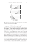
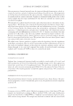
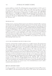
EFFECT OF Gp4G ON SKIN TISSUES 477 known to play key roles in many cellular signaling pathways involved in hair follicle biol- ogy. Previous studies showed that VER is capable of blocking the interaction between cells and other ECM molecules, such as collagen I, fi bronectin, and laminin. Pericellular matrix expansion also requires interactions among VER, hyaluronan, and CD44 (36,37). This macromolecular complex increases the viscoelastic nature of the pericellular matrix, creating a highly malleable extracellular environment that controls cell shape, a necessary step for cell proliferation, migration, and histogenesis (38–42). As expected, VER is expressed in larger amounts in tissues undergoing rapid develop- ment or remodeling (42,43). Previous studies have shown that during embryogenesis, a short fragment (839 bp) of human VER is suffi cient to promote hair follicle formation (44). In fact, involvement of chondroitin sulfate proteoglycans, such as VER, in hair fol- licle development and/or hair growth has been suggested because of their dermal papilla (DP)-specific localization in anagenic hair follicles (32). Although the functional roles of VER in hair follicle development and hair growth is not completely understood, several re- searchers have suggested that VER functions as an inhibitor of cell–cell or cell–extracel- lular matrix adhesion, allowing DP-cell aggregation (45,46). In the present study, VER was highly expressed in the matrix and in the cytoplasm of dermal papillar cells (Figure 6) that are in the anagen phase, confi rming previous results (32,38). Ten follicles of all subtypes were imaged, and the staining was quantifi ed in each section. Importantly, the immunostaining for VER in the Gp4G–treated group was 78% higher than in the control (Figure 6c). The anagen phase of the hair follicle is character- ized by intense proliferation of both connective tissue cells and papillar cells (5,29,38). As indicated above, the cell proliferation, which is indicative of anagen, is accompanied by increasing expression of VER that signals quiescent papillar cells to restart a new growth cycle (29,38). Accordingly, increasing VER expression in the Gp4G formulation-treated group indicates that Gp4G formulation activates the papillar cell population, sustaining the anagen phase and the consequent hair elongation. Analysis of Figures 2–6 clearly shows that the Gp4G formulation provides a general acti- vation of dermal tissues. Although all components present in the Gp4G formulation may collectively help to obtain this effect, the effect of antioxidant agents and red pepper ex- tract cannot solely explain the results obtained (23,47). The key ingredient present in the Gp4G formulation, which is Gp4G itself, has not yet been linked to the mechanisms by Figure 6. Effect of Gp4G formulation on versican synthesis. (a) Immunoreaction for versican in the bulbic dorsal anagen follicles. (b) Immunoreactions after color isolation of the specifi c versican immunostaining. (c) Mean density (in arbitrary units) obtained in the immunocytochemical analysis for versican in the bulb dorsal follicles measured after Image J treatment to isolate pixels from the VER primary antibody stained.

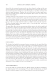
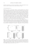
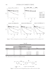
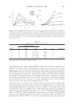
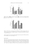
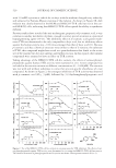
JOURNAL OF COSMETIC SCIENCE 478 which the compound acts in the cells and, therefore, a series of experiments was per- formed to propose a hypothesis for the cellular mechanism of Gp4G. CELLULAR MECHANISM OF Gp4G Experiments aiming to test the effect of Gp4G on cellular metabolism were conducted using immortalized epithelial Hela cells and primary fi broblasts from Swiss mouse embryos. Although Gp4G may be interacting with a variety of cell types including fol- licular stem cells, the aim of these studies is to provide a general intracellular activation mechanism. The other ingredients present in the Gp4G formulation were not included in order to unveil the cellular role of Gp4G alone. By evaluating changes in absorption in the wavelength region of nucleotide absorption, we were able to quantify the increase in the nucleotide concentrations inside cells as a function of Gp4G concentration. Gp4G enters Hela cells and the uptake follows a saturation plateau (Figure 7). At 6 μM (5 ppm), Gp4G increases cell viability and the number of cell colonies by 28% and 13%, respec- tively (Figure 7b,c), confi rming that Gp4G stimulates Hela cells in culture. Similar, but more pronounced, results were obtained with primary fi broblasts (Figure 7d). Gp4G suffers asymmetric enzymatic cleavage to produce GTP and GMP in CAS (13–15, 48). Recent data showed that this conversion also happens in human cells (49). Cells have a variety of enzymes that link the total nucleoside phosphate pool with ATP-bioenerget- ics. For example, nucleoside diphosphate kinases, which catalyze transfer of γ-phosphate from nucleoside 5′-triphophates to nucleoside 5′-diphosphates and adenylate kinases, Figure 7. Gp4G internalization in epithelial cells, viability, and colony growth. Hela cell uptake of Gp4G. (a) Measurements were obtained by light absorption at 258 nm in Hela cells after disruption with SDS. Ab- sorptions were discounted from control values. (b) MTT assay of cell viabilities for 105 Hela cells after 3-hr incubation with Gp4G in DMEM. (c) Number of colonies of Hela cells after 8 days in growth under Gp4G at 6μM (5 ppm). (d) MTT assay of cell viabilities for 105 primary fi broblast cells after 3-hr incubation with Gp4G in DMEM. * Signifi cantly different from controls, p 0.05.
Purchased for the exclusive use of nofirst nolast (unknown)
From: SCC Media Library & Resource Center (library.scconline.org)