EFFECT OF Gp4G ON SKIN TISSUES 473 experimental groups were simultaneously incubated with DAB, and the reaction was im- mediately interrupted with PBS after a specifi c period (1–5 min, depending on the anti- body). After immunostaining, the sections were counterstained with Mayer’s hematoxylin (Merck, Darmstadt, Germany). For each immunoreaction, the negative control was performed by replacing the primary antibodies with the respective non-immune serum at similar concentrations or by omitting the primary antibody step from the protocol. Sections were examined with a Nikon Eclipse E600 microscope. Images were captured using a digital camera (COOL SNAP-PROcf color) and Image-Pro Plus software (Media Cybernetics, Silver Spring, MD). IN VITRO CELL PROLIFERATION, VIABILITY, AND METABOLISM OF Gp4G Hela and primary fi broblasts from Swiss mouse embryos were grown in DMEM containing 100 mg/ml of streptomycin and 100 UI/ml of penicillin at 37°C, in a humid atmosphere with 5% CO2. The cell uptake of Gp4G and the viability assays were conducted with extract from CAS (from FarmaService BioExtract) and with Gp4G acquired from Sigma-Aldrich. Only the results from the CAS extract are shown in the text, although the results obtained were basically the same. Gp4G from both sources were dissolved in DMEM at 5–10 ppm concentrations and sterilized by 0.22-μm fi lters to incubate the cells. Cell viability as- says were conducted by trypan blue uptake and MTT (3-(4,5-dimethylthiazol-2-yl)- 2,5-diphenyltetrazolium). Only cells with more than 90% viability were used in all experiments. After three hours of incubation, MTT assays were used according to the literature, in order to measure the cell viability. Briefl y, cells were incubated with 2 mg/ml MTT in PBS for two hours. DMSO was used to dissolve the formazan, and light absorption at 550 nm was read in a Shimadzu UV3901-PC spectrophotometer or in a Tecan-Infi nite M200 reader plate. For the colony growth assay, Hela cells were grown in six-well plates with 1000 cells/ well over eight days. After most colonies reached more than 105 cells, they were counted, fi xed with 10% formic aldehyde, and stained with 1.0% violet crystal. The experiment for determination of ATP concentrations was conducted using biolumi- nescence and a luciferin-luciferase assay and expressed in 105 cells, which were counted using the classical hematocytometer technique (25). Other nucleotides were measured in six-well plates with 1 × 10⁶ cells/well. Cells with Gp4G at 5 ppm and the control cells were incubated for three hours. After that, the media was removed and 1 ml of 0.8 M HCIO4 was added (chemical lysing and mechanically detaching the cells) to denature all enzymes, thus interrupting all enzymatically catalyzed reactions that consume or produce ATP, besides all others that could affect nucleotide concentrations. After 15 minutes over ice, 1 ml of KOH at 0.8 M in aqueous solution was added. The supernatant was carefully removed and transferred to a Millipore fi ltration unit for exclusion of molecules larger than 5 kDa by centrifugation at 12,000g and 4°C. Finally, 20 μl of the fi ltered samples (containing the nucleotides) was either submitted to the bioluminescent assay or injected in a Shimadzu 20A HPLC system. A column of anionic exchange, Shimadzu WAX-IT (0.46 × 5 cm), was used in a linear gradient fl ow of 1 ml/min, starting with 20 mM of phosphate buffer at pH 7.0, and changed after 30 minutes to a 480 mM phosphate buffer at pH 6.0. External standards for ADP, AMP, GTP, GDP, GMP, and Gp4G were used to identify and quantify the concentrations of all these nucleotides in the cell extracts. A biological triplicate for experiment and control was used to calculate the average and standard deviation (error bars in the graphs).

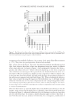
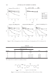
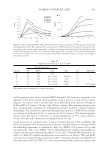
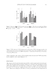
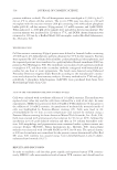
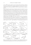
JOURNAL OF COSMETIC SCIENCE 474 RESULTS AND DISCUSSION HAIR GROWTH AND SHIFT IN THE FOLLICLE CYCLE The fi rst experimental evidence that the Gp4G formulation favors hair growth was ob- tained by comparing the size of hair shafts of Wistar rats that were (experiment) or were not (control) treated with the Gp4G formulation for an average of four weeks, during a hair follicle cycle induced by depilation. The experiment was repeated twice on a total of 20 animals. Hair shafts were on the average 20 ± 2% longer (Figure 1a). Rats have two basic types of hair: guard or overcoat, which can be further sub-grouped into monotrichs, awl, and auchenes and undercoat hairs, which are also called zigzags. Separating the hair into two size classes, i.e., above 10 cm (overcoat hairs) and below 6 cm (mostly undercoat hairs), we observed that there was an increase in the size of both undercoat (12 ± 5%) and overcoat (27 ± 2%) hair shafts (Figure 1b,c). However, the increase observed in the un- dercoat shafts was smaller than the increase observed in the overcoat shafts and was not statistically signifi cant. The most conspicuous effect of the Gp4G formulation was ob- served in the overcoat shafts, which may be the fi rst indication of a possible shift in the hair cycle. By counting papillar cells in the bulb of ten follicles in each section for a total of three sections, we were also able to show an average increase in the papillar cell count (32 ± 4%) in the bulbs of the treated follicles (Figure 1d). Therefore, the follicles are somehow being stimulated to induce hair growth. Similar effects have been shown to oc- cur in Wistar rats treated with Hibiscus rosa-sinensis extracts. The action mechanism of Hibiscus seems to be related to the effect of β-catenin, which prolongs the anagen phase and/or shortens the telogen phase (7,12). Recent results indicate that a seed extract of Hibiscus abelmoschus favors FGF-2 activity, which may also help to explain the result cited above (23). In order to understand the effect of Gp4G in hair follicles, histological follicles that were either in anagen or telogen phases were counted (Figure 2) (24,26). This method is based on the fact that the medulla of telogen follicles receives a different staining in longitudi- nal sections at the height of the sebaceous glands (see Methods section). Treatment with Gp4G formulation causes a decrease in the number of telogen follicles and an increase in the number of anagen follicles when compared with the control group. This provides strong evidence that the average increase in the hair length was promoted by a shift in the hair cycle, either by an expansion in the anagen phase or a shortening of the telogen phase. In terms of potential application to humans, we do not expect to see an increase in Figure 1. Effect of Gp4G formulation treatment on the hair follicle structure. (a) Average length of hair shafts, length of (b) undercoat and (c) overcoat hair shafts, and (d) papillar cell count in the bulb of follicles. Cell count represents an average number for all types of follicles. Signifi cantly different from controls, * p 0.05.
Purchased for the exclusive use of nofirst nolast (unknown)
From: SCC Media Library & Resource Center (library.scconline.org)