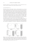
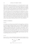
STABILITY OF FERULIC ACID 503 APPENDIX V: SPECTROSCOPIC DATA OF PVG AND ITS DIMER Compound 1: 4-vinylguaiacol. Colorless to pale yellow oil nature of TLC was the same as that of a standard 4-vinylguaiacol sample. IR spectra indicate that the molecule has –Ar, –OH, C–O, and C=C groups. Mass spectra (MS) show molecular ion M- as 149, which indicates that the molecular formula is C9H10O2. This compound was identifi ed as 4-vinylguaiacol IR cm-1: 3600 (–OH), 3100, 3050, 3000, 2980, 2900 (–Ar), 1620 (C=C), 1280, 1050 (C–O), 930, 790 MS (m/z): 149 (M-), 134 1 H NMR (CDCl3, 300 MHz) δ: 3.92 (3H, s, MeO), 5.12 (1H, dd, Ha), 5.57 (1H, s, OH), 5.62 (1H, dd, Hb), 6.64 (1H, dd, Hx), 6.86-6.94 (3H, m, Ar-H) (6.78 1H, d, Ar-H 6.86 1H, dd, Ar-H 7.03 1H, d, Ar-H) UV (methanol): λmax = 220nm, 280nm. Compound 2: 1, 3-bis (4-hyroxy-3-methoxyphenyl) -1-butene. Compound 2 was white acerate crystal, which was crystallized slowly by standing. It could be easily oxidized in air and could not be recrystallized in the usual solvents. The IR spectrum bore a strong resemblance to that of 4-vinylguaiacol except that, notably, the band at 970 cm-1 was weaker with respect to the band at 1036 cm-1. IR cm-1: 3600 (–OH), 3100, 3050, 3000, 2980, 2900 (–Ar), 1620 (C=C), 1280, 1050 (C–O), 930, 790 MS(m/z): 299 (M-). Mo- lecular formula: C18H20O4 1 H NMR (CD3COCD3, 300 MHz) δ: 1.19-1.40 (3H, d, Me), 3.52 (1H, m, Hx), 3.83 (6H, s, MeO), 6.28 (1H, dd, Hb), 6.32 (1H, d, Ha), 6.74-7.04 (6H, m, Ar-H) UV (methanol): λmax = 270nm. Compound 1: PVG. Compound 2: Dimer of PVG.

Purchased for the exclusive use of nofirst nolast (unknown)
From: SCC Media Library & Resource Center (library.scconline.org)