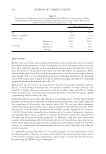
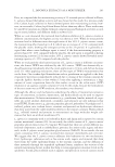
MENOPAUSE INCREASES FERRITIN IN SKIN 179 chelators on skin (12) in which we showed the protective effects of a UVA-photo activated iron chelator, our results indicate that increased iron as a result of menopause may make postmenopausal skin more susceptible to chronological aging as well as photo aging in postmenopausal women. Reductions in TAC and VEGF also support increased vulnera- bility to oxidative stress and reduced wound healing, respectively, as a result of increased ferritin in skin. Finally, we observed levels of ferritin in NHEK comparable to that of HepG2 cells, a liver cancer cell that is rich in iron. Taken together, our study shows that menopause increases ferritin in skin and that this increased iron is a potential risk factor contributing to the manifestation of accelerated skin aging after menopause. REFERENCES ( 1) S. Stevenson and J. Thornton, Effect of estrogens on skin aging and the potential role of SERMs, Clin. Interv. Aging, 2, 283–297 (2007). ( 2) S. Verdier-Sevrain, F. Bonte, and B. Gilchrest, Biology of estrogens in skin: Implications for skin aging, Exp. Dermatol., 15, 83–94 (2006). ( 3) M. A. Ali, A. Akhmedkhanov, A. Zeleniuch-Jaquotte, P. Toniolo, K. Frenkel, and X. Huang, Reliabil- ity of serum iron, ferritin, nitrite, and association with risk of renal cancer in women, Cancer Det. Prev., 27, 116–121 (2003). ( 4) Q. Yang, J. Jian, S. Katz, S. B. Abramson, and X. Huang, 17β-estradiol inhibits iron hormone hepcidin through an estrogen responsive element half-site, Endocrinology, 153, 1370–1378 (2012). (5) J. Jian, E. Pelle, and X. Huang, Iron and menopause: Does increased iron affect the health of postmeno- pausal women? Antioxid Redox Signal, 11, 2939–2943 (2009). (6) C. Pourzand, R. D. Watkin, J. E. Brown, and R. M. Tyrell, Ultraviolet A radiation induces immediate release of iron in human primary skin fi broblasts: The role of ferritin, Proc. Natl. Acad. Sci. USA, 96, 6751–6756 (1999). (7) J. Jian, E. Pelle, Q. Yang, N. Pernodet, D. Maes, and X. Huang, Iron sensitizes keratinocytes and fi bro- blasts to UVA-mediated matrix metalloproteinase-1 through TNF-alpha and ERK activation, Exp. Dermatol., 20, 249–254 (2011). (8) L. M. Milstone, B. D. Adams, J. Zhou, V. L. Bruegel Sanchez, and J. Shofner, Stratum-specifi c expres- sion of human transferring receptor increases iron in mouse epidermis, J. Invest. Dermatol., 126, 648– 652 (2006). (9) M. Hickey, S. R. Davis, and D. W. Sturdee, Treatment of menopausal symptoms: What shall we do now? Lancet, 366, 409–421 (2005). (10) D. Peus, R. A. Vasa, A. Meves, M. Pott, A. Beyerle, K. Squillace, and M. R. Pittelkow, H2O2 is an important mediator of UVB-induced EGF-receptor phosphorylation in cultured keratinocytes, J. Invest. Dermatol., 110, 966–971 (1998). (11) E. Pelle, X. Huang, T. Mammone, K. Marenus, D. Maes, and K. Frenkel, Ultraviolet-B-induced oxida- tive DNA base damage in primary normal human epidermal keratinocytes and inhibition by a hydroxyl radical scavenger, J. Invest. Dermatol., 121, 177–183 (2003). (12) E. Pelle, J. Jian, L. Declercq, K. Dong, Q. Yang, C. Pourzand, D. Maes, N. Pernodet, D. B. Yarosh, and X. Huang, Protection against ultraviolet A-induced oxidative damage in normal human epidermal ke- ratinocytes under post-menopausal conditions by an ultraviolet A-activated caged-iron chelator: A pilot study, Photodermatol. Photoimmunol. Photomed., 27, 231–235 (2011).

Purchased for the exclusive use of nofirst nolast (unknown)
From: SCC Media Library & Resource Center (library.scconline.org)