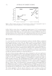
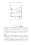
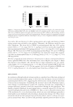
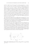
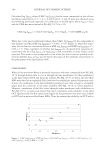
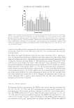
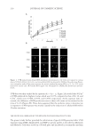
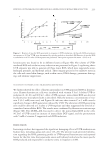
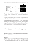
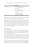
KINETICS OF UVB IRRADIATION-MEDIATED ROS GENERATION IN HUMAN KERATINOCYTES 215 in response to UVB could be detected as early as 15 min (data not shown), and the explanation of such prompt effect may be due to the reducing property of vitamin C. Vitamin C was shown to reduce and maintain function of glutathione in human keratinocytes (38). Even though further investigations as well as in vivo studies are necessary to support these fi ndings in human keratinocytes and results observed in this study may not necessarily translate into effects observed in vivo in human skin. The present study provided the ini- tial evaluating method supplanting the use of animal for the evaluation of cosmetic and cosmeceutical constituents. Also, the knowledge gained from the present study might benefi t the development of sunscreen products and help to design appropriate active in- gredients that effectively block the dominant oxidative species induced by UVB. CONCLUSIONS In conclusion, we revealed herein the kinetic of ROS generation and identifi ed specifi c ROS in UVB-exposed human skin keratinocytes. These data enhance the understanding on skin biology and benefi t the development of strategies in protection of UVB-mediated damages. ACKNOWLEDGMENT This research was supported by Cell-based Drug and Health Products Development Re- search Unit, Chulalongkorn University and Agricultural Research Development Agency. The authors thank Mr. Krich Rajprasit for editing the manuscript. REFERENCES (1) W. Guo, Y. An, L. Jiang, C. Geng, and L. Zhong, The protective effects of hydroxytyrosol against UVB- induced DNA damage in HaCaT cells, Phytother. Res., 24, 352–359 (2010). (2) C. Matito, N. Agell, S. Sanchez-Tena, J. L. Torres, and M. Cascante, Protective effects of structurally diverse grape procyanidin against UV-induced cell damage and cell death, J. Agric. Food Chem., 59, 4489–4495 (2011). Figure 6. Effect of vitamin C on UVB-mediated oxidative stress in keratinocytes. Cells were treated with 0.5, 1, and 5 mM of vitamin C for 1 h before 90 J/m2 UVB irradiation. (A) ROS level was determined by DCFH2-DA probe using fl uorescence microplate reader. Data point represent the mean ± SD (n = 4), *p 0.05 versus UVB-treated cells. (B) Cellular ROS signal were determined by DCFH2-DA fl uorescence measurements.

JOURNAL OF COSMETIC SCIENCE 216 (3) G. J. Fisher, S. C. Datta, H. S. Talwar, Z. Q. Wang, J. Varani, S. Kang, and J. J. Voorhees, Molecu- lar basis of sun-induced premature skin aging and retinoid antagonism, Nature, 379, 335–339 (1996). (4) K. H. Kraemer, Sunlight and skin cancer: another link revealed, Proc. Nat. Acad. Sci. USA, 94(1), 11–14 (1997). (5) I. G. Kirkinezos, and C. T. Moraes, Reactive oxygen species and mitochondrial diseases, Cell Dev. Bio., 12, 449–457 (2001). (6) Y. Liu, G. Fiskum, and D. Schubert, Generation of reactive oxygen species by mitochondrial electron transport chain, J. Neurochem., 80(5), 780–787 (2002). (7) Y. Chen, E. Mcmillan-ward, J. Kong, S. J. Israels, and S. B. Gibson, Mitochondrial electron-transport- chain inhibitors of complexes I and II induce autophagic cell death mediated by reactive oxygen species, J. Cell Sci., 120, 4155–4166 (2007). (8) B. Halliwell, and M. C. Gutteridge, Oxygen toxicity, oxygen radicals, transition metals and disease, Biochem. J., 219, 1–14 (1984). (9) T. P. A. Devasagayam, J. C. Tilak, K. K. Boloor, K. S. Sane, S. S. Ghaskadbi, and R. D. Lele, Free radi- cals and antioxidants in human health: current status and future prospects, J. Assoc. Phys. India, 52, 794–804 (2004). (10) R. I. Salganik, The benefi ts and hazards of antioxidants: controlling apoptosis and other protective mechanisms in cancer patients and the human population, J. Am. Coll. Nutr., 20(5), 464–472 (2001). (11) O. Bossi, M. Gartsbein, M. Leitges, T. Kuroki, S. Grossman, and T. Tennenbaum, UV irradiation in- creases ROS production via PKC delta signaling in primary murine fi broblasts, J. Cell Biochem., 105(1), 194–207 (2008). (12) V. J. Thannickal, and B. L. Fanburg, Reactive oxygen species in cell signaling, Am. J. Physiol. Lung Cell Mol. Physiol., 279, 1005–1028 (2000). (13) Y. Saitoh, A. Miyanishi, H. Mizuno, S. Kato, H. Aoshima, K. Kokubo, and N. Miwa, Super-highly hydroxylated fullerene derivative protects human keratinocytes from UV-induced cell injuries together with the decreases in intracellular ROS generation and D NA damages, J. Photochem. Photobiol. B. Biol., 102, 69–76 (2011). (14) H. Wang, and I. E. Kochevar, Involvement of UVB-induced reactive oxygen species in TGF-β biosyn- thesis and activation in keratinocytes, Free Radical Bio. Med., 38, 890–897 (2005). (15) A. E. Maccubin, J. Przybyszewski, M. S. Evans, E. E. Budzinski, H. B. Patrzyc, M. Kulesz-Martin, and H. C. Box, DNA damage in UVB-irradiated keratinocytes, Carcinogenesis, 16(7), 1659–1660 (1995). (16) S. Lisby, R. Gniadecki, and H. C. Wulf, UV-induced DNA damage in human keratinocytes: quantita- tion and correlation with long-term survival, Exp. Dermatol., 14(5), 349–355 (2005). (17) H. Wang, and J. A. Joseph, Quantifying cellular oxidative stress by dichlorofl uorescein assay using microplate reader, Free Radical Bio. Med., 27, 612–616 (1999). (18) G. Ana, F. Eduarda, and L. L. F. C. Jose, Fluorescence probes used for detection of reactive oxygen spe- cies, J. Biochem. Biophys. Methods, 65, 45–80 (2005). (19) V. P. Bindokas, J. Jordan, C. C. Lee, and R. J. Miller, Superoxide production in rat hippocampal neu- rons: selective imaging with hydroethidine, J. Neurosci., 16, 1324–1326 (1996). (20) L. Benov, L. Sztejnberg, and I. Fridovich, Critical evaluation of the use of hydroethidine as a measure of superoxide anion radical, Free Radical Bio. Med., 25, 826–831 (1998). (21) M. A. Barbacanne, J. P. Souchard, B. Darblade, J. P. Iliou, F. Nepveu, B. Pipy, F. Bayard, and J. F. Arnal, Detection of superoxide anion released extracellularly by endothelial cells using cytochrome c reduction, ESR, fl uorescence and lucigenin-enhanced chemiluminescence techniques, Free Radical Bio. Med., 29, 388–396 (2000). (22) K. Setsukinai, Y. Urano, K. Kakinuma, H. J. Majima, and T. Nagano, Development of novel fl uores- cence probes that can reliably detect reactive oxygen species and distinguish specifi cs species, J. Biol. Chem., 278, 3170–3175 (2003). (23) J. G. Mohanty, J. S. Jaffe, E. S. Shulman, and D. G. Raible, A highly sensitive fl uorescent micro-assay of H2O2 release from activated human leukocytes using a dihydroxyphenoxazine derivate, J. Immunol. Methods, 202, 133–141 (1997). (24) M. Zhou, Z. Diwu, N. Panchuk-Voloshina, and R. P. Haugland, A stable nonfl uorescent derivative of resorufi n for the fl uorometric determination of trace hydrogen peroxide: applications in detecting the activity of phagocyte NADPH oxidase and other oxidases, Anal. Biochem., 253, 162–168 (1997).
Purchased for the exclusive use of nofirst nolast (unknown)
From: SCC Media Library & Resource Center (library.scconline.org)