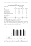
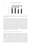
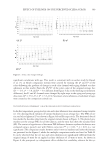
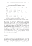
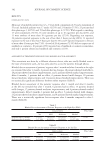
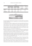
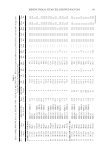
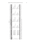
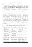
Table III Gender and Age Differences in the Melanocyte and Melanoblast Populations/0.1 mm2Interfollicular Epidermis of the Skin Treated With MEL/NB-UVB Alone and MEL/NB-UVB +MTK Freq/Cell no MEL/NB-UVB, MTK No. of melanocytes/0.1 mm2 No. of melanoblasts/0.1 mm2 (A)Gender Female Male Female Male Freq MEL/NB-UVB alone 0% (0/6) 9.1% (1/11) 0% (0/6) 9.1% (1/11) MEL/NB-UVB +MTK 55.6% (5/9) 62.5% (5/8) 55.6% (5/9) 62.5% (5/8) Cell no MEL/NB-UVB alone 0 (N =6) 0.22 ± 0.22 (N =11) 0 (N =6) 0.46 ± 0.46 (N =11) MEL/NB-UVB +MTK 19.7 ± 11.2 (N =9) 32.9 ± 12.3 (N =8) 22.2 ± 10.4 (N =9) 33.5 ± 12.5 (N =8) (B)Age Young Old Young Old Freq MEL/NB-UVB alone 12.5% (1/8) 0% (0/9) 12.5% (1/8) 0% (0/9) MEL/NB-UVB +MTK 62.5% (5/8) 55.6% (5/9) 62.5% (5/8) 55.6% (5/9) Cell no MEL/NB-UVB alone 0 (N =8) 0.27 ± 0.27 (N =9) 0 (N =8) 0.57 ± 0.57 (N =9) MEL/NB-UVB +MTK 30.0 ± 12.7 (N =8) 22.3 ± 11.1 (N =9) 36.4 ± 12.5 (N =8) 19.6 ± 10.0 (N =9) Freq, frequency of differentiation of melanocytes/melanoblasts after the treatment with MEL/NB-UVB alone and MEL/NB-UVB +MTK. MTK, treatment with FFC, collagen peptide, elastin peptide, and UCB-MSCCS. Young patient, from 4 to 30 years Old patient, from 31 to 76 years. Each datum of cell number (Cell no) is an average ± standard error of the mean. No significant differences in gender and age were observed. 399 MESENCHYMAL STEM CELL-DERIVED FACTORS

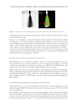
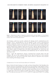
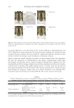
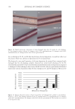
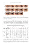
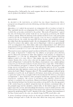
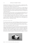
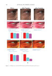
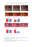
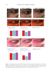
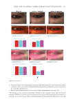
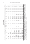
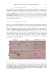
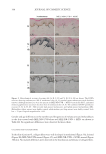
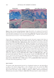
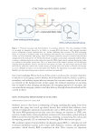
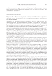
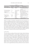
400 JOURNAL OF COSMETIC SCIENCE Elastin fibers in lesional (Figure 3F) skin were greatly decreased compared with those in nonlesional (Figure 3E) skin. The elastin fibers were torn and sparse. However, in the skin treated with MEL/NB-UVB +MTK (Figure 3H), elastin fibers were greatly increased. In the skin treated with MEL/NB-UVB +MTK, elastin fibers were increased in all cases (100%, 17/17) even in the treated skin after only 8 days (Table IC, patient 38). Elastin fibers became thick and encircled the rete ridge. However, in the skin treated with MEL/NB-UVB alone, no recovery of elastin fibers was observed (Figure 3G, Table IA). By contrast, in the skin treated with MEL/NB-UVB +MT, increase in elastin fibers was observed in 14 of 17 patients (82.4%, Table II). This difference in the recovery rate of elastin fibers between MEL/NB-UVB alone and MEL/NB-UVB +MT was statistically significant (p 0.001), whereas the difference between MEL/NB-UVB +MTK and MEL/NB-UVB +MT was not statistically significant. Moreover, in the patients treated with MEL/NB-UVB +MT (29, 33, and 34), no recovery of elastin fibers and no differentiation of melanocytes and melanoblasts was observed. Difference in the frequency of complete recovery of elastin fibers between MEL/NB-UVB +MTK (100%, 17/17) and MEL/NB-UVB +MT (42.9%, 6/14) was statistically significant (p 0.01). DISCUSSION The present study demonstrated that the loss of epidermal melanocytes and melanoblasts as well as the reduced elastin fibers in the vitiliginous skin was restored by MEL/ NB-UVB exposures and MTK irrespective of gender and age. Even in patient 49, who received only three sessions of MEL in addition to MTK, melanocyte/melanoblast differentiation was induced. However, MEL/NB-UVB alone failed to induce melanocyte/ Figure 3. Azan staining revealed well-developed collagen fibers (Coll) in the nonlesional (A), lesional (B), MEL/NB-UVB-treated (C), and MEL/NB-UVB +MTK–treated (D) skin in a similar density. By contrast, elastin fibers (Elas) were greatly decreased in the lesional (F) and MEL/NB-UVB–treated (G) skin compared with nonlesional skin (E). However, in the skin treated with MEL/NB-UVB +MTK (H), elastin fibers were dramatically increased. Scale bar =25 µm. Original magnification ×400.
Purchased for the exclusive use of nofirst nolast (unknown)
From: SCC Media Library & Resource Center (library.scconline.org)