146 JOURNAL OF COSMETIC SCIENCE
was assayed for an amino acid distribution by means of a fully automatic NEXERA XR
system amino analyzer (Shimadzu, Kyoto, Japan) equipped with the separation column
(150mm x 3.0mm) packed with 3.0 µm diameter YMC-Triart C18 beads (YMC Co., LTD,
Kyoto, Japan). Solution A (10 mM Na
2 HPO
4 ,10 mM Na
2 B
4 O
7 ,pH 8.2) and B (acetonitrile
:MeOH :H
2 O =45 :45 :10 v/v) were used as a mobile phase.
QUANTIFICATION OF THIOL RESIDUE OF KERATIN
The thiol residue was quantified according to the method adapted by Obata et al.16 A
mixture of the keratin (1 wt%), chicoric acid (4.0 mM) and copper (II) chlorophyllin (4 ppm)
was dissolved in phosphate buffer (0.1 M, pH 7.4) and warmed at 40°C for 12 hours. The
resulting reaction mixture was diluted by a factor of 10× with PBS and dialyzed against
the same buffer at 4°C for 12 hours using a membrane tubing (molecular weight cut-off of
14.0 kDa). Subsequently, the dialyzed solution (0.4mL) was mixed with an ethanol solution
of DTNP (0.5 mM, 0.1mL) and warmed at 37°C for 30 minutes, then was subjected to the
absorption measurement at 386 nm using a U-3310 UV-spectrometer (Hitachi, Ibaraki,
Japan). The number of thiol residue in keratin protein was calculated with the calibration
curve, which was obtained using L-cysteine as a reference compound viz. the cysteine
concentration was well correlated to the absorbance at 386 nm by equation (absorbance at
386 nm) =cysteine concentration (μM) × 0.0092 +0.3144.
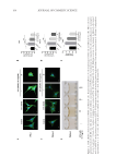
FLUORESCENCE MICROSCOPIC OBSERVATIONS OF FITC-I DYEING HAIR SAMPLE
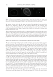
The fluorescence measurement was carried out according to the procedure reported by
Nakamura et al.17 Several strands of hairs were added into carbonate buffer (0.1 M, pH 5.0)
containing FITC-I (180 ppm) and left for 12 hours. The resulting hair fibers were washed
three times with 0.5mL of carbonate buffer (0.1 M, pH 9.5) and sliced cross-sectionally by
means of a microtome to obtain the specimens (about 20 µm in thickness), then subjected
to the fluorescent measurement at excitation wavelength of 470–495 nm and fluorescence
wavelength of 510–550 nm using a Olympus BX51 fluorescence microscope (Olympus
Corporation, Tokyo, Japan) equipped with a U-MNIBA3 mirror unit (Olympus Corporation,
Tokyo, Japan). The shutter speed was 2.5 seconds and the ISO sensitivity was 800.
OPTICAL MICROSCOPIC OBSERVATIONS FOR EXAMINATION OF POLYPHENOL-TREATMENT OF
BLEACHED HAIR
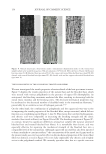
Biological microscopes (Olympus Photomax with the proper attachments) were used for
bright field observations (Olympus Corporation, Tokyo, Japan). The Olympus objective
lenses were: (bright field) Plan ×10, PlanApo ×20 and PlanApo ×40 (Olympus Corporation,
Tokyo, Japan). Original photo tube accessories were modified to adapt the digital camera
that was automatically controlled by a desktop computer to optimize for lighting, ISO
levels and focusing measures. The images in JPEG and RAW formats were developed by
means of Lightroom ver.3 and Photoshop Elements ver. 9 software (Adobe Inc., San Jose,
CA). Image enhancement included color level correction, noise reduction, and contrast and
brightness adjustments the purpose of the enhancement was to make the image appear
nearly identical to that seen actually by the observer.
was assayed for an amino acid distribution by means of a fully automatic NEXERA XR
system amino analyzer (Shimadzu, Kyoto, Japan) equipped with the separation column
(150mm x 3.0mm) packed with 3.0 µm diameter YMC-Triart C18 beads (YMC Co., LTD,
Kyoto, Japan). Solution A (10 mM Na
2 HPO
4 ,10 mM Na
2 B
4 O
7 ,pH 8.2) and B (acetonitrile
:MeOH :H
2 O =45 :45 :10 v/v) were used as a mobile phase.
QUANTIFICATION OF THIOL RESIDUE OF KERATIN
The thiol residue was quantified according to the method adapted by Obata et al.16 A
mixture of the keratin (1 wt%), chicoric acid (4.0 mM) and copper (II) chlorophyllin (4 ppm)
was dissolved in phosphate buffer (0.1 M, pH 7.4) and warmed at 40°C for 12 hours. The
resulting reaction mixture was diluted by a factor of 10× with PBS and dialyzed against
the same buffer at 4°C for 12 hours using a membrane tubing (molecular weight cut-off of
14.0 kDa). Subsequently, the dialyzed solution (0.4mL) was mixed with an ethanol solution
of DTNP (0.5 mM, 0.1mL) and warmed at 37°C for 30 minutes, then was subjected to the
absorption measurement at 386 nm using a U-3310 UV-spectrometer (Hitachi, Ibaraki,
Japan). The number of thiol residue in keratin protein was calculated with the calibration
curve, which was obtained using L-cysteine as a reference compound viz. the cysteine
concentration was well correlated to the absorbance at 386 nm by equation (absorbance at
386 nm) =cysteine concentration (μM) × 0.0092 +0.3144.
FLUORESCENCE MICROSCOPIC OBSERVATIONS OF FITC-I DYEING HAIR SAMPLE
The fluorescence measurement was carried out according to the procedure reported by
Nakamura et al.17 Several strands of hairs were added into carbonate buffer (0.1 M, pH 5.0)
containing FITC-I (180 ppm) and left for 12 hours. The resulting hair fibers were washed
three times with 0.5mL of carbonate buffer (0.1 M, pH 9.5) and sliced cross-sectionally by
means of a microtome to obtain the specimens (about 20 µm in thickness), then subjected
to the fluorescent measurement at excitation wavelength of 470–495 nm and fluorescence
wavelength of 510–550 nm using a Olympus BX51 fluorescence microscope (Olympus
Corporation, Tokyo, Japan) equipped with a U-MNIBA3 mirror unit (Olympus Corporation,
Tokyo, Japan). The shutter speed was 2.5 seconds and the ISO sensitivity was 800.
OPTICAL MICROSCOPIC OBSERVATIONS FOR EXAMINATION OF POLYPHENOL-TREATMENT OF
BLEACHED HAIR
Biological microscopes (Olympus Photomax with the proper attachments) were used for
bright field observations (Olympus Corporation, Tokyo, Japan). The Olympus objective
lenses were: (bright field) Plan ×10, PlanApo ×20 and PlanApo ×40 (Olympus Corporation,
Tokyo, Japan). Original photo tube accessories were modified to adapt the digital camera
that was automatically controlled by a desktop computer to optimize for lighting, ISO
levels and focusing measures. The images in JPEG and RAW formats were developed by
means of Lightroom ver.3 and Photoshop Elements ver. 9 software (Adobe Inc., San Jose,
CA). Image enhancement included color level correction, noise reduction, and contrast and
brightness adjustments the purpose of the enhancement was to make the image appear
nearly identical to that seen actually by the observer.