178 JOURNAL OF COSMETIC SCIENCE
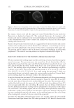
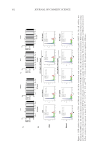
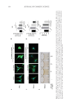
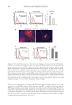
Figure 3. PLE induces DNA repair signals γH2AX and p53 in epidermal keratinocytes independently of
DNA damage. (A) Representative flow cytometry profiles showing levels of γH2AX (solid magenta line) in
primary human keratinocytes, untreated or treated with PLE for 8 days. As a positive control of γH2AX
induction, a separate sample of cells was treated for 24 hours with 1 µM doxorubicin (dotted red line). Dotted
black line is negative isotype antibody control (B) quantitation of the percent of γH2AX-positive cells
according to the gate shown in A (C) Western blot for p53 expression in cells untreated or treated with PLE
for 8 days. Cells treated with Doxorubicin as in A were included as a control for the induction of p53. GAPDH
is used as protein-loading control. Bottom histogram represents the optical density normalized to GAPDH
and relative to control (CT) (D) immunofluorescence for expression of p53 (arrows) of cells treated for 5 days
as indicated. Scale bar: 50 µm. (E) representative nuclear image of comet assay analyses of human epidermal
keratinocytes untreated or treated with PLE for 6 days, as in A and B, to test for chromosomal fragmentation
and (F) quantitation of DNA fragmentation measured by tail length, relative to untreated (UN) control cells
(n =299/302). Scale bar: 10 µm. Data are mean ± SD of duplicate samples representative of two independent
experiments. *p 0.05 ns: not significant (p 0.05).
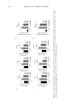
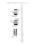
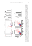
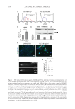
Figure 3. PLE induces DNA repair signals γH2AX and p53 in epidermal keratinocytes independently of
DNA damage. (A) Representative flow cytometry profiles showing levels of γH2AX (solid magenta line) in
primary human keratinocytes, untreated or treated with PLE for 8 days. As a positive control of γH2AX
induction, a separate sample of cells was treated for 24 hours with 1 µM doxorubicin (dotted red line). Dotted
black line is negative isotype antibody control (B) quantitation of the percent of γH2AX-positive cells
according to the gate shown in A (C) Western blot for p53 expression in cells untreated or treated with PLE
for 8 days. Cells treated with Doxorubicin as in A were included as a control for the induction of p53. GAPDH
is used as protein-loading control. Bottom histogram represents the optical density normalized to GAPDH
and relative to control (CT) (D) immunofluorescence for expression of p53 (arrows) of cells treated for 5 days
as indicated. Scale bar: 50 µm. (E) representative nuclear image of comet assay analyses of human epidermal
keratinocytes untreated or treated with PLE for 6 days, as in A and B, to test for chromosomal fragmentation
and (F) quantitation of DNA fragmentation measured by tail length, relative to untreated (UN) control cells
(n =299/302). Scale bar: 10 µm. Data are mean ± SD of duplicate samples representative of two independent
experiments. *p 0.05 ns: not significant (p 0.05).