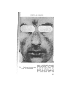
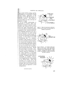
COSMETICS AND DERMATITIS By E. J. MOYNAHAN, M.R.C.P.* •?!?MAXKIND HAS used cosmetics since }•,!i:i :' ear y palaeolithic times, and although !•: we cannot be certain that the con- ?i' nection between health and beauty i•!i: dates back so far, we know that there ii!. ! has been a dose relationship between i? medicine and the cosmetic art for a :::•1ong time. It is of some interest 5:-:that, among the earliest medical ?,:writings, we find several cosmetic :'.recipes in the Ebers Papyrus, and ,:-many references to such preparations - are to be found in the works of the early alchemists and herbalists. Until .. recently medicine has approached the problem of dermatitis empirically, and it is only witlain the present :. century that the scientific founda- tions for its study have been laid. :.• It had long been known that cos- metics produce reactions in the skin, but there was no scientific proof of ß : this until Kesten and Lazlo showed, in 1931, that nineteen out of twenty- one cases of dermatitis, due to cos- metics, gave positive patch tests to one or more ingredients in the preparation used. We owe a great deal to Sulzberger and his colleagues, who performed no fewer than ten thousand tests 6n a thousand sub- jects, and threw considerable light ß Assistant Physician, Dermatological Department, Guy's Hospital, London. on the commoner causes of trouble with cosmetics.. Very. few primary irritants are used in modern cosmetics: these include permanent waving agents, which attack the keratin fibres and break the disulphide linkages and other chemical bonds between adja- cent keratin molecules depilatories, with a closely related chemical action high concentrations of alkali, which reduce the sulphur in the disulphide hnks and rupture them in the same way and the oxidation hair dyes, such as paraphenylenediamine. Most cases of dermatitis produced by cosmetics are due to sensitisation to some substance in the preparation. It should be emphasised that such cases are extremely few, considering the large number of people who use cosmetics daily, year in, 'year out, without any ill effect on their skin. It speaks well for those engaged in the production and manufacture of these preparations that so little trouble follows their use. We doc- tors have not got such good records in this respect, -because a fair number of the preparations we use in the treatment of skin disease produce dermatitis, and some of the newer chemotherapeutic agents, such as sulphonamides and antibiotics (par- 203

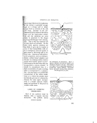
JOURNAL OF THE SOCIETY OF COSMETIC C, HEMISTS ticularly penicillin), are notorious for the ease with which they produce sensitisation after local application to the skin. It is important, in view of what follows, to distinguish between a primary irritant and a sensitising agent. Primary irritants are sub- stances which, when applie•d in sufficient concentration to the skin of a normal individual, will provoke an inflammatory reaction. A sensitis- ing agent, on the other hand, can. be applied with impunity to the skin of most individuals without ill effect, but on the skin of a susceptible person, will give rise to a dermatitis. Before going into details it would be useful here to give a brief account of the microscopic structure of the skin. The skin is composed of two parts, the epidermis and the dermis. The epidermis arises from the basal layer from which all the overlying layers are derived. This layer rests on the dermis, from which it is separated by a very thin basement membrane. It is anchored to the dermis by certain processes which project from inside the basal cells. These are fibrous in nature but their exact composition remains unknown. Above the basal layer we find several layers of cells known as prickle cells because of the presence of inter- connecting fibrils between adjacent cells. These fibrils appear to run through several cells and skirt the nucleus of each. They help to keep the cells together and some authori- ties believe that they are the pre- cursors of keratin. As we ascend 204 . the prickle layer towards the sur- face of the skin, the cells tend to flatten out and granules accumulate in their cytoplasm. This layer, which may be two or three cells thick, is known as the granular layer, but is not present everywhere in the skin. It is absent, for example, in the eyelids. In certain parts of the skin, such as the palms and soles, there is another layer to be found in the epidermis between the granu- ß lar layer and the horny layer. The cells composing this layer contain clear granules, and it is known as the stratum lucidum. The horny layer is composed of flat squames with a fibrous protein, keratin, forming a shell or rind round an inner kernel of lipids. The presence of an intact horny layer ensures protection against chemical, mechanical, radia- tion and other injuries and provides also a defence against bacterial and fungal attack. The sebum, secreted:i:.i by the sebaceous glands which open !i:• into the hair follicles, also helps protect the underlying living cells" :: against these irritants. , .: The dermis itself contains nerves, blood vessels and'supporting.i! } tissues. The vessels in the upper, papillary, part of the dermis play prominent part in the inflammatorY.i:!ii response of the skin which we knoW? as dermatitis. Thus, when an irri2?i tant penetrates the protective keratini:':?i or horny layer, the living cells irritated and a reaction is seen in the skin which varies from redness (dU• to dilatation of these vessels), witll:?i} associated subjective itching, gross swelling and even the produc?7•
Purchased for the exclusive use of nofirst nolast (unknown)
From: SCC Media Library & Resource Center (library.scconline.org)