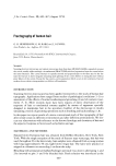
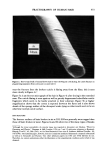
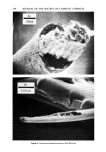
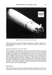
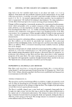
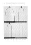
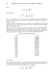
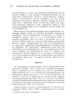
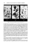
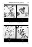
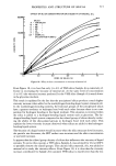
450 JOURNAL OF THE SOCIETY OF COSMETIC CHEMISTS end of each interval. Hairs fractured in solvents were soaked a minimum of 16 hr before Instron testing. Variation in pH was accomplished by addition of acetic acid or ammonium hydroxide. It was noted after 16 hr that the pH of these systems was not af- fected by the hair. Relative humidities other than 50 -+ 1% (room condition) were ob- tained for 20, 71, 79 and 90% by using saturated salt systems (9) in capped jars containing sample hanging racks and a fixed post. Samples to be fractured were equili- brated for a minimum of three days at 21 m iøC in these chambers. A glycerin/water mixture equivalent to a water activity of 50% RH was prepared using 22.5% water and 77.5 % anhydrous glycerin by weight. Stress/strain measurements on hairs were made on a Model TT-B Instron Tester. Hairs were mounted in special holders of stainless steel with chamfered holes and secured with tapered Teflon plugs. The working length was 12.7 mm and most experiments were done at a constant rate of extension of 200%/min. Some determinations were made at one-tenth this speed (20%/min). Hairs run in the humidity chambers were fixed between small screw clamps to provide the desired working length and suspended between a hook on the Instron and the fixed post in the chamber. The hook extended down through a small hole in the cover. The screw clamp subjected the hair to a 2- to 3~g stress during the conditioning period which was ignored since the resulting extension is negligibly small. The hairs were viewed in a Coates and Welter Cwik Scan Model 100-2 Field Emission Scanning Electron Microscope. Prior to viewing, the hairs were sputter coated with gold/palladium in a Denton DV-515 Evaporator equipped with a Model DSM-1 Sput- tering Module. The sputtering was done at 10 mA and 150 millitorr for 1.5 min in in- tervals of 30 sec. For SEM viewing the hairs were mounted perpendicular to the surface of a standard Coates and Welter specimen stub. The length of hair exposed below the fracture varied but was generally of the order of 1 mm. Both sides of the fracture were examined in all cases. The optical microscopy was carried out on hairs immersed in a dish of water on the stage of a Zeiss Universal Photomicroscope. The hairs were mounted on a Hoffmann hose clamp modified to permit use of the screw-driven bearings to stretch the specimen. RESULTS WET FRACTURE The fracture surfaces of virgin hairs broken under water are often quite flat as shown in Figure 1. Here it appears that the fracture started at a small zone on the edge of the fiber and radiated from there in a plane perpendicular to the axis. Such detailed evi- dence of fracture propagation is seldom seen but, in general, the new surface is remarkably devoid of evidence of the cellular and subcellular fibrous structure of hair the fracture pattern is much more similar to the brittle fractures of glass or carbon (10) than to the fracture patterns of other natural fibers such as cotton (11). The cuticle fracture is also planar and perpendicular to the fiber axis and again evidence of cellular structure is lacking there is little sign of delamination or axial slippage of the

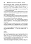
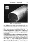
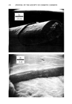
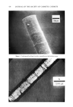
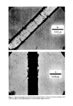
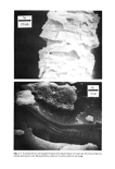
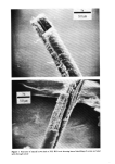
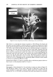
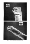
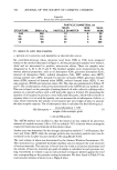
FRACTOGRAPHY OF HUMAN HAIR 451 25m Figure 1. Fracture of natural brown hair in water cuticle cells. At about a quarter of the fiber diameter from the fracture plane it can be seen that the cuticle has cracked circumferentially this too is typical of the wet fracture pattern. In Figure 1 the break fits Brown and Swift's description (7) "almost as if cut with a knife." In a few cases, however, we observed wet fractures like that in Figure 2a where there is a gross mismatch of the fracture planes of the cuticle and cortex. A gap between cuticle and cortex can be seen in Figure 2b. Figure 2c is the opposing fracture surface which is complementary to the first. Here there is much clearer evidence of another circumferential failure of the cuticle far from the fracture surface. Such frac- tures of the cuticle were frequently observed several diameters from the fracture sur- face, as seen in Figure 3. Although such cuticle fractures might have occurred at the instant of failure, the more intriguing possibility is that the cuticle fails well before the cortex. Two types of experi- ment proved that this is indeed the case. First, hairs that were extended in water to just short of failure, relaxed in water and then dried and examined in the SEM showed many cracks like those in Figure 3. Second, when hairs were extended incrementally in water and examined in the polarizing microscope it was evident in some cases, as shown in Figure 4, that at least part of the cuticle had ruptured. In Figure 4a the rup- ture lines are clearly seen in top focus against the bright corticle matter between slightly uncrossed polars. In Figure 4b at edge focus in the same view it can be seen that
Purchased for the exclusive use of nofirst nolast (unknown)
From: SCC Media Library & Resource Center (library.scconline.org)