j. Soc. Cosmet. Chem., 38, 77-82 (March/April 1987) Measurement of the rate of hair growth using a fluorescent tracer technique JONATHAN R. MATIAS, RHODA ALANI, and NORMAN ORENTREICH, Biomedical Research Station, Orentreich Foundation for the Advancement of Science, RD 2, Box 3 70A, Cold Spring, NY 10516 (J.R.M., R.A., N.O.), Berg Institute, New York University Medical Center, 550 First Avenue, New York, NY 10016 (J.R.M.), and Department of Dermatology, New York University School of iMedicine, 550 First Avenue, New York, NY 10016 (N.O.). Received September 25, 1986. Synopsis The rate of hair growth was measured in the guinea pig by determining the distance between the fluores- cent bands produced by subcutaneous injections of sodium fluorescein. The minimal dose required to produce a visible fluorescent band was determined to be 30 mg/kg for the guinea pig. Rate of hair growth may also be measured in localized areas of the skin using intradermal injections at dosages as low as 0.25 mg. This method is simple, inexpensive, accurate, and less time-consuming than radiotracer methods. INTRODUCTION The measurement of the rate of hair growth is important in the evaluation of hormonal, cosmetic and drug effects on hair growth. Over the years a number of methods have been developed in an attempt to measure this parameter accurately. Notable among these are the capillary method by Saitoh et al. (1), the photographic technique by Burgess and Edwards (2), or the 35-S radioautographic method by Harkhess and Bern (3). The major disadvantages of the first two methods are that: (a) it is difficult to measure the rate of growth of the same hairs over a period of time, and (b) they cannot be easily adapted for hair growth measurements in animals with dense hair coat. These problems may be avoided by the injection of radiolabeled cystine which is incorporated into the hair shaft during the growth phase or anagen cycle. This method has been used successfully to measure rate of growth in sheep (3), guinea pigs (4), rats, and human beings (5). However, this technique is less convenient for the routine evaluation of drugs which might influence the rate of hair growth because of: (a) the prohibitive cost of isotope procurement and disposal, and (b) the long waiting time before the radioau- tographs may be developed. Fluorescein binds readily to the epidermal cells of the skin. Since this chemical may be 77

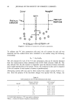
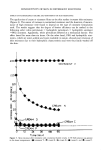
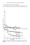
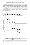
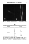
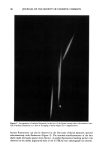
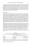
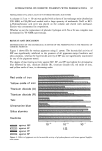
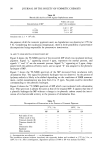
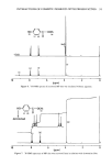
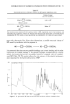
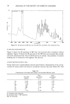
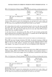
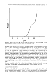
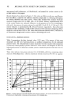
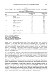
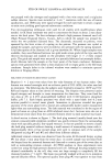
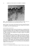
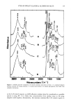
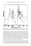
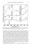
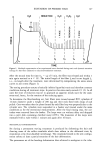
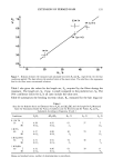
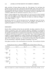
78 JOURNAL OF THE SOCIETY OF COSMETIC CHEMISTS incorporated into the hair shaft during the growth phase of the hair cycle, it should be possible to utilize fluorescein as a marker for the growing hair. This report examines the usefulness of fluorescent tracers as an approach to accurately determine the rate of hair growth. MATERIALS AND METHODS Adult male Hartley guinea pigs (250-300 g) were purchased from Camm Laboratory Animals, Inc. (Wayne, NJ). Male Syrian golden hamsters (10-12 weeks) were ob- tained from Harlan-Sprague Dawley (Farmersberg, VA). The animals were maintained at a photoperiod of 12-h light and 12-h dark. Food and water were provided ad libitum. An area 3 to 4 cm on the back of the animals was plucked manually. Three days after epilation, various doses (3 to 60 mg/kg body weight) of sodium fluorescein (Aldrich Chemical Co., Milwaukee, WI), dissolved in 0.2 cc of physiological saline, were in- jected subcutaneously on alternate days at a site distant from the epilated area. The hairs were plucked three days after the last injection, rinsed with hexane, and dry- mounted on glass slides. The incorporation of this dye into the hairs of hamsters was also evaluated using subcutaneous injections of sodium fluorescein. In another experi- ment, various doses (0.05 to 1.0 mg) of sodium fluorescein in 0.1 cc of saline were injected intradermally into epilated dorsal skin of the guinea pig. The hair of the guinea pig was classified according to the method of Dawson (6). The fluorescent bands in I and type II hair were visualized using a fluorescence microscope (Ortholux II, Leitz) equipped with a drawing attachment and integrated with a com- puterized graphics calculator (Numonics Corp., Lansdale, PA). Type I hairs are long (10-25 mm in length) and broad, with the apex ending abruptly in a sharp point. Type II hairs are shorter than type I and characterized by a very fine, wool-like apical portion. Linear growth was determined by measuring the distances between the center of two fluorescent bands. RESULTS Figures la and lb show the presence of fluorescent bands along the shafts of type I and II hairs of the guinea pig when examined under the fluorescence microscope. Although autofluorescence was present along the entire length of the shaft, the amount of fluores- cein incorporated after subcutaneous administration at a dose of 30 mg/kg was sufficient to provide a distinct fluorescent band. Some of the nonspecific fluorescence, possibly due to lipids coating the hair shaft, can be eliminated with chloroform or ether. The effects of various doses of subcutaneously injected sodium fluorescein on the inten- sity of the band in guinea pig hair are shown in Table I. Doses of 15 mg/kg and below produced barely detectable banding or no fluorescence at all. The animals tolerated the treatments well and untoward effects were not observed even at the highest dose. So- dium fluorescein was also administered intradermally at doses ranging from 0.05 mg to 1.0 mg/injection. Fluorescent bands were present even at the lowest dose (0.05 mg). However, a greater fluorescence intensity, suitable for growth measurements, was ob- served at a dose of 0.25 mg and higher.
Purchased for the exclusive use of nofirst nolast (unknown)
From: SCC Media Library & Resource Center (library.scconline.org)