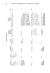
J. Soc. Cosmet. Chem., 38, 247-262 (July/August 1987) A quantitative diffuse reflectance method using Fourier transform infrared spectroscopy for determining siloxane deposition on keratin surfaces HELEN M. KLIMISCH and GRETCHEN S. KOHL, Dow Corning Corporation, 2200 West Salzburg Road, Midland, MI 48640 and JOAN M. SABOURIN, Delta College, University Center, Auburn, MI 48710. Received December 1 O, 1986. Synopsis Methodology was developed for the detection and quantitation of siloxanes on keratin fibers using DRIFTS. Sample preparation procedures were defined and optimized for surface and bulk analysis of the fiber. The effort has demonstrated that DRIFTS band ratio data can be correlated to mg/kg Si data generated independently by atomic absorption analysis of hair fibers. A linear relationship was observed over the 250 to 1840 mg/kg Si range of this study. The deposition of any material that has a distinctive IR band could also be assayed by this method. The success of the method is almost totally dependent upon sample preparation and generation of a KBr-reflective surface to produce the surface analysis. INTRODUCTION Keratin fibers, such as human hair, undergo environmental damage due to UV radia- tion and atmospheric oxidation, mechanical damage by grooming devices, and chemical damage from permanent waving lotions, bleaches, dyes, and even shampoos. So-called conditioning agents can be used on the hair, usually after shampooing, to produce improvements in fiber properties by depositing ingredients on the fiber surface. Some typical organic conditioning agents are organic quaternary ammonium compounds and long-chain animal proteins. Previous studies (1-3) have shown that polysiloxanes, especially organofunctional polysiloxanes, also act as conditioning agents. Silicone de- position on hair fibers has been investigated using electron microscopy, atomic absorp- tion (AA), and electron spectroscopy for chemical analysis (ESCA). Scanning electron microscopy (SEM) provides a qualitative means for detecting the presence of a film on the fiber surface. ESCA is a surface analysis technique ø capable of determining the con- centration of elemental silicon within the top 50A of the fiber. An AA method was developed (4) for quantitatively measuring the silicon content of the whole fiber. The general conclusion based on these methods is that combing ease or conditioning improved as the silicon content of the fiber increased. AA provides information on 247

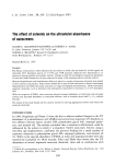
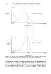
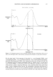
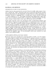
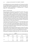
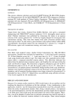
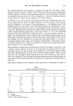
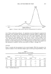
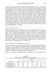
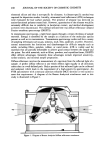
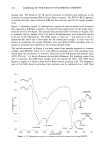
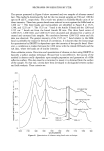
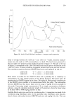
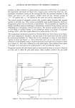
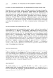
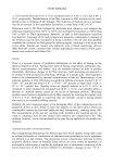
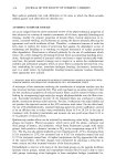
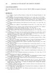
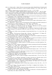
248 JOURNAL OF THE SOCIETY OF COSMETIC CHEMISTS elemental silicon and thus is non-specific for siloxanes. A siloxane-specific method was required for deposition studies. Initially, attenuated total reflectance (ATR) techniques were evaluated for hair surface analysis. The presence of siloxane was detected on amine-functional polymer treated hair. However, quantitation of the siloxane would be extremely difficult due to variability in hair/prism contact, and method development activities were discontinued. The method selected here was diffuse reflectance infrared Fourier transform spectroscopy (DRIFTS). In transmission spectroscopy, a light beam passes through a certain thickness of sample material. Energy is absorbed by the sample as a function of the molecular species present as well as its concentration. Transmission spectroscopy works well for a variety of sampling techniques such as gas, solution, mull, film, sandwich, or pellet. However, normal transmission has limitations and disadvantages for difficult samples such as solids, including fillers, powders, rubber, or cured resins. ATR is widely used for materials that are partially deformable to achieve good contact between the sample and the prism. For solid materials, such as fillers, powders, and crystalline forms, DRIFTS offers definite advantages. Generally these advantages include improved sensitivity, surface analysis, and minimal sample preparation. Diffuse reflectance involves the measurement of a spectrum from the reflected light of a sample. A perfect diffuse reflector is one which reflects light equally in all directions rather than in a well-defined path. Only a portion of this reflected light can be collected and analyzed, which leads to the requirement of a high-sensitivity spectrometer. An FTIR spectrometer with a mercury cadmium telluride (MCT) detector is well suited to meet this requirement. A diagram of the Barnes Analytical attachment used in this study is illustrated in Figure 1. m4 or sample m6 m• Figure 1. Diagram of Barnes Analytical diffuse reflectance attachment.
Purchased for the exclusive use of nofirst nolast (unknown)
From: SCC Media Library & Resource Center (library.scconline.org)