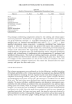
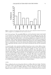
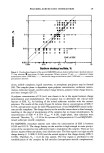
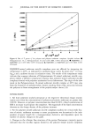
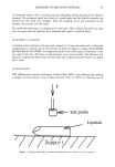
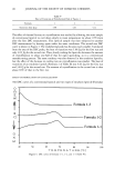
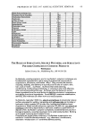
PREPRINTS OF THE 1997 ANNUAL SCIENTIFIC SEMINAR 53 Table 2: Symptoms related to groups of causes Symptoms groups of reddening tighteness spots stinging patches of :::::::::::::::akin:::::::::::::::::::::::: causes scurf ::: :::: CONTACT 44 :::: 37:: i::: 7 21 ::: I 8 4 5 : ß LIFESTYLE 38 1 32::: I 0 2 0 2 CYCLE 0 0 •19::: :::: 0 0 0 0 0 ENVIRON 130ß 33 ::::: i :: 5 12 ::!5::: I 5 2 A new set of variables describing symptoms related to groups of causes was subsequently constructed for the 197 women was grouped e.g. as reddening reddening due to causes related to environmental conditions, contact or lifestyle. A multiple correspondence analysis was then performed on these data and 3 associations were found. Association I pooled spots due to menstrual cycle and spots and reddening due to causes related to life style association 2 combined reddening, itching, tightness and stinging due to causes related to contact association 3 pooled reddening, tightness, stinging, patches of scuff and flaking skin due to causes related to environmental conditions. When perceived symptoms of skin sensitivity were related to clinical parameters and biophysical properties several links were found. For example, the symptom of reddening due to causes related to contact was found to be associated with an increased transepidermal water loss. By contrast, reddening due to causes related to life style or environmental conditions was associated with an increased blushing response to tactile stimuli and an elevated a* (red axis) value by Chromameter measurement. Although these results are as yet tentative, they suggest that self-attributed sensitive skin is a multi-dimensional phenomenon. The confirmation of the associations of symptoms described above on a larger sample with a wider range of ages will allow better characterization of groups of individuals, with different types of self-attributed sensitivity. Further studies will be necessary to confirm the potential relationship between self-attributed skin sensitivity and objective clinical, biophysical and biological parameters. THE EFFECTS OF AN ALPHA HYDROXY ACID (GLYCOLIC AcIo) ON HAIRLESS GUINEA PIG SKIN H. L. Hood', M. E. K. Kraeling', M. G. RobP and R. L. Bronaugh' •Office of Cosmetics and Colors, 2Office of Beltsville Technical Operations, Food & Drug Administration, Washington, DC Introduction Since repeated daily use of alpha hydroxy acids (AHAs) have been demonstrated to alter the structure of the stratum comeurn • (SC), viable epidermis and dermis 2, it seemed possible that use of AHAs could result in skin barrier alterations that may effect changes in the absorption of topically applied chemicals. To test this theory, hairless guinea pigs (HGPs) were treated with AHA formulations according to a protocol determined to increase SC turnover. A commercial moisturizing lotion was used as the control. Following treatment, HGPs were sacrificed for in vitro measurement of the percutaneous absorption of hydroquinone (HQ) and musk xylol. Methods Estimation of Skin Cell Renewal Time The dansyl chloride (DC) staining technique 3 was used to measure SC renewal time. The dye was triturated at 5% (w/w) into petrolatum and applied to the backs of HGPs for 24 hr. An [IV lamp was employed to visualize the stain. In measuring SC renewal time after pretreatment with the control or AHA lotions, application of the lotions was continued until DC fluorescence disappeared. In Vivo Topical Exposure Studies The test lotions used for the topical exposure studies consisted of an oil-in-water emulsion containing 5% glycolic acid (GA) or 10% GA at pH 3.0. The control was Vaseline Intensive Care

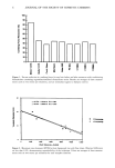
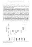
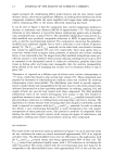
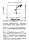
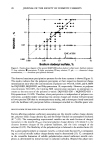
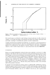
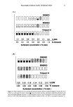
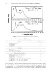
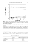
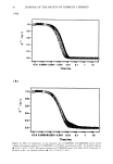
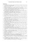
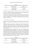
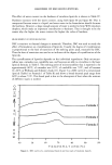
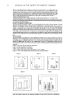
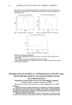
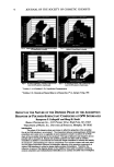
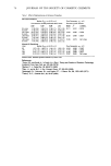
54 JOURNAL OF THE SOCIETY OF COSMETIC CHEMISTS (vIc) Lotion for Sensitive Dry Skin (Chesebrough-Ponds, USA), pH 7.8. Lotions were applied once daily at 3 mg/cm 2 for 3 weeks (Sundays excluded) to designated sites on the back of HGPs. At the end of 3 weeks, HGPs were sacrificed for the in vitro percutaneous absorption studies. Percutaneous Absorption In vitro skin absorption of HQ and musk xylol was measured through HGP skin dermatomed to a thickness of-200-gm and assembled in flow-through diffusion cells. Skin viability was maintained in the diffusion cells during the 24-hr studies by using physiological media. The barrier integrity of untreated, GA- and VIC-treated HGP skin was assessed by determining [3H]water absorption prior to each study (20 rain test•). []4C]HQ or []4C]musk xylol was applied to skin in an oil-in-water emulsion vehicle at a 2% or 0.20% concentration, respectively. At 24 hr, unabsorbed compound was washed from skin with soap and water and the radioactivity recovered in the skin and receptor fluid was determined. Histology Full-thickness skin samples were taken from treatment sites, fixed in 10% formalin, and then stained for light microscopic evaluation. The number of epidermal cell layers was determined from fixed skin sections the thickness of the viable epidermis was measured from photographs (50X magnification). Results and Discussion The effect of GA formulations on skin mover was determined by measuring the disappearance of DC from the skin surface. HGPs were able to tolerate a once-a-day dosing with the GA formulations for 2 weeks, followed by DC application, and then continued daily dosing until fluorescence disappeared. The 5% and 10% GA treatment reduced SC turnover times by 36% and 39%, respectively, compared to the VIC control (Fig. 1). A 3-week pretreatment time was, Fig 2. Glycollc Acid Effect on Stretum Corneum Turnover • 25 e, 20 I-- i• 15 • lO I-- 5 t/ntreltld vllemlne IC s• olycollc Acid 10'/. Olycollc Acid Pretreatment FormulatlonB therefore, sufficient to effect an increase in SC turnover in HGPs used for the skin absorption studies. The effect of the various pretreatment formulations on the barrier integrity of HGP skin was determined by measuring [3H]water absorption through skin prior to the HQ and musk xylol in vitro studies. No significant difference in the percent of the applied [•H]water absorbed was detected between untreated skin and the pretreatment formulations. The percutaneous absorption of HQ was also unaffected by pretreatment applications. No significant difference was found between the partial (receptor fluid or skin) or total absorption values measured for the different treatment groups (Table 1). In general, musk xylol absorption was unaffected by pretreatment applications. However, total absorption values for VIC, 5% GA, and 10% GA were significantly different from untreated skin but were not different from each other. Table 1. In Vitro Skin Absorption of Two Cosmetic Ingredients Following Pretreatment of Hairless Guinea Pig Skin Pretreatment Formulations Untreated VI_•C 5% GA 10%(•A -Q receptor fluid 4.25 + 0.56 5.89 + 0.30 6.44 + 0.88 4.40 4- 0.52 skin 15.0 + 0.67 13.2 + 1.07 15.4 + 1.02 16.1 + 1.82 total 19.3 + 0.43 19.0 + 1.03 21.8 4- 1.87 20.5 + 2.10 Musk Xylol receptor fluid 30.3 + 2.49 23.6 + 0.17 20.7+ 4.23 21.6 + 3.56 skin 18.0 + 2.55 18.8 + 1.21 16.1 + 3.20 18.8 4- 2.12 total 48.3 + 1.73 42.4 4- 1.07 36.7 + 1.31 40.4 + 1.72 Although no increase in penetration of HQ or musk xylol was observed following pretreatment with GA lotions to HGP skin, the structure of skin was drastically altered. Histological examination of the skin revealed a similarity in thickness of untreated and VIC- treated skin (5.0 cell layers) and a thickening of the epidermis in skin treated with 5% GA (9.8 cell layers) and 10% GA (8.1 cell layers). Measurements of the viable epidermal thickness
Purchased for the exclusive use of nofirst nolast (unknown)
From: SCC Media Library & Resource Center (library.scconline.org)