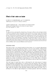
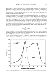
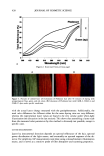
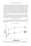
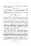
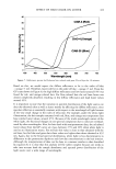
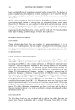
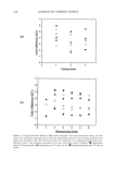
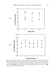
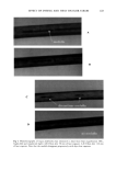
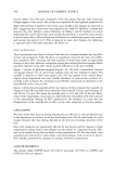
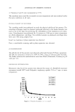
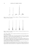
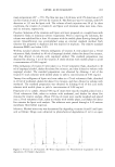
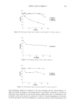
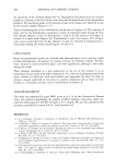
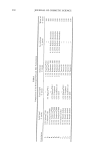
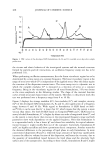
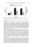
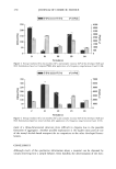
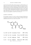
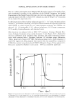
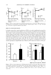
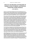
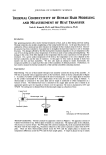
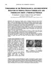
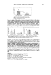
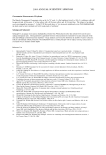
500 JOURNAL OF COSMETIC SCIENCE COMPARISON OF THE BIOMECHANICAL AND BIOSYNTHETIC BEHAVIOR OF NORMAL HUMAN IrIBROBLASTS AND ]:IBROBLASTS FROM A FOREHEAD WRINKLE M. Jouandeaud ', C. Viennet 2, S. Bordes', B. C!oss •, and P. Humbert 2 •R&D Department. SILAB, Brive, France 2Lab of Engineering and Cutaneous Biology, University Hospital St. Jacques, Besancon, France Introduction. With increasing age, metabolic activity of fibroblasts decreases and they lose their adhesion capacity to collagen fibers, thereby limiting the possibility of organizing dermal tissue •. Johnson et al. 2 sho•ved that fibroblasts obtained from donors of different ages presented a general deceleration in term of protein synthesis. Moreover, fibroblasts aged artificially by successive passages synthesized lower quantities of type I and III collagens than young fibroblasts 3'n'5. The behavior of old fibroblasts has already been investigated, but fe•v studies were carried out on the functional capacities of old fibroblasts directly resulting from a •vrinkle. We thus examined the behavior of human fibroblasts obtained from biopsies from a forehead •vrinkle. The aim of this study •vas to investigate the biomechanical and biosynthetic behavior of fibroblasts from •vrinkles and to compare them to those of normal fibroblasts. Collagen I, the major collagen of the dermal matrix, was chosen as a nmrker for the study of the synthetic activity of fibroblasts. The biomechanical behavior of fibroblasts front wrinkles •vas studied using a three-dimensional model of collagen gels to •Ssualize reorganization capacities. We then examined the possibility of a plant extract to restore and compensate losses of the synthesis and org•mization capacities of a collagen gel by fibroblasts from •vrinkles. We thus sho•ved that selected soy peptides could improve the synthesis and contractile capacities of fibroblasts from wrinkles. Contractile capacity of fibroblasts front wrinkles is reduced The biomechanical behavior of hmnan fibroblasts from •vrinkles and that of normal fibroblasts was studied using a model of human fibroblast cultures suspended in a collagen gel. Normal huntan fibroblasts have considerable properties of reorganization and reorientation of the collagen fibers 6. Fibroblasts influence the alignment of collagen fibers, which in turn affects the distribution of fibroblasts 7. These properties are manifested physically in the lattices by contraction of the gel. This phenomenon is primarily related to locomotion of the cells inside collagen lattices 6. It was sho• that normal human fibroblasts suspended in a collagen gel could contract and reorg•mize collagen fibers, in contrast to fibroblasts from wrinkles that exlfibited significant reduced contractile capacities (-66%) (fig. D. i - ...... ,•,,...•: ß •..•' . .... ß Figure 1: Compafimn of the biomech•ic• behavior of nom• fibroblasts and basic fibrobl• offices after 3 days in a collagen la•ice. Collagen I synthesis is reduced in fibroblasts from wrinkles. We determined the synthesis capacities of fibroblasts obtained from biopsies of forehead wrinkles and normal human fibroblasts. In order to determine ff collagen production by fibroblasts from wrinkles and normal fibroblasts •vere different, •ve quantified collagen synthesis by the fibroblasts cultivated in monolayers. The collagen I synthesis capacity of fibroblasts frown •vrinkles was lcnver (-70%) than that of normal fibroblasts (fig. 2).

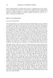
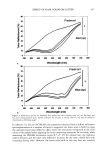
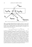
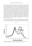
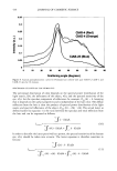
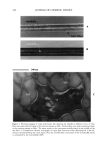
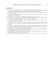
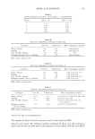
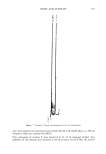
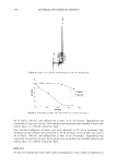
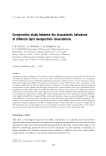
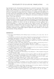
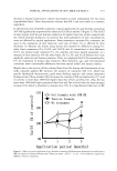
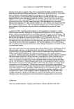
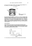
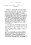
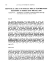
2004 ANNUAL SCIENTIFIC SEMINAR 501 Normal hum•m fibroi•sts F•broblasta I•n• wri•kles * SignScant result according to Student's rest •0. 05) •gure 2: Comp•mn of •e me•bolic activity of noel fib•bi•Ls and fibrobiasLs from •nkles, Study of •oy pepff•s w•h the potent&l of •proving the bioscharted behavior and the •ynthe• eapae•es of fibrobla• from •ink•s. We ex•fined •e possibili• of restoring •e con•a•le •d synCeric capadfies of fibroblasts •om writes by u•ng • a•ive in•edient obtained •er s•ee•ng several mw materiffs. •e beM•or of noir fibrobl•ts •d •ose from writes in te•s of collagen I syn•esis •d con•cfile c•adties w• sm•ed in •e pr•ence of a selected soy peptides in comparison to 'vim•n C, a reference molecule known to sfimtfiate •e metabolism of m•y derm• proteins. It w• found •at sdected soy pepfi&s were most effective for promot•g collagen I •n•esis •d e•c•g •e con•acfile c•acity of hum• fibroblasts from •e base of writes. 25 Figures 3a & 3b: Effect of selected soy peptides on collagen I synthesis and on the contractile capacity of normal human fibroblasts or those t¾om wrinkles. Comparison with vitamin C (1 pg.mL4). Conclusion. It was thus of interest to further understand the link between tissue modification and cell functiol• The data we have presented suggest the existence of differences in biomechanical behavior and synthesis capacities between normal fibroblasts and fibroblasts from the base of wrinkles. Changes of cell metabolism appear to be one of the keys of mechanical degradation of mature skin, manifested on the cutaneous surface by the appearance of wrinkles. Limiting the appearance of wrinkles thus involves boosting the synthesis of derreal proteins such as collagens I and III, but also restoring the adhesion and contractile capacities of fibroblasts to the extracellular matrix to favor their biomechanical properties which can again ensure their role of tissue support. In the present work, we tested a untobet of potential active ingredients and found that selected soy peptides concomitantly restored the synthesis of proteins such as type I collagen of by fibroblasts from wrinkles and the reorganization capacities of collagen fibers of these cells. This effect could be compared to vitamin C. In order to further understand the modifications of cell functions with aging, we plan to use normal human fibreblasts and those from wrinkles obtained from the same donor_ This could show if cell modifications are progressive or appear suddenly by including donors of different ages. References. 1 Piischel HU et al. d. Photochern. Photobiol. B. 27, 39-6 (1995) 2 Johnson BD et al. Lab. Invest., 55, 490-96 (1986) 3 Varani Jet al. d. Investig. Dermatol. Syrnp. Proc. 3, 41-4 (1998) 4 Varani Jetal. Am. d. Patho. 158, 931-42 (2001) 5 Chung JH et al. Photoderrnatol. Photo•rnrnunol. Photomeal. 12, 84-9 (1986) 6 Ehrlich HP et al, Tissue Cell. 22, 407-17 (1990) 7 Tada A et al. 22 • IFSCC congress Edinburgh Proc., 1 (2002)
Purchased for the exclusive use of nofirst nolast (unknown)
From: SCC Media Library & Resource Center (library.scconline.org)